Article • Digital pathology
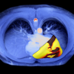
From single-tumour to pan-cancer AI diagnostics
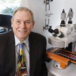
AI is transforming the way cancer is diagnosed and treated. Dr Yuri Tolkach shared his group's advances in developing AI-based tools for oncological pathology at the DPAI Europe congress.