Article • Molecular meets digital
Single-cell multiplex imaging: a powerful tool for digital pathology
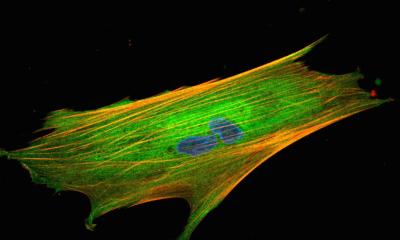
Multiplex imaging can play a critical role in unravelling the tumour microenvironment. The potential and benefits of the emerging approach – a way to extract information from human tissue samples by visualising many more biomarkers than traditional microscopy – was highlighted in presentations during the 36th European Congress of Pathology in Florence, Italy. Speakers also discussed novel technologies to transform clinical care and deep learning for pathology biomarkers for precision oncology.
Report: Mark Nicholls

In his presentation, Dr Teijo Pellinen indicated that understanding the diverse cell states and molecular composition within the tumour microenvironment is crucial for improving risk stratification and predicting therapy responses. The Senior University Researcher at the Institute for Molecular Medicine Finland (FIMM) at the University of Helsinki, Finland, said multiplex tissue imaging is ‘a great tool’ for discovering ‘clinically meaningful associations’ and can answer different scientific questions.
Explaining the digital image processing and analysis steps in multiplex tissue profiling, he discussed different spatial tissue imaging approaches, from IHC-based systems, immunofluorescence, mass spectrometry and tyramide signal amplification systems. ‘Some are more simple and high-throughput, meaning that they can cover large numbers of samples, while others are more suited to deep biological profiling and can cover up to 100 protein targets but are typically limited to fewer sample numbers with expensive infrastructure costs,’ said Pellinen. ‘Basically, the choice of multiplexing system depends on the biological and scientific questions.’
A more advanced way of doing image analysis
Pellinen highlighted how his FIMM group has developed an in-house approach that combines tryamide signal amplification and cyclic immunofluorescence in an affordable high-throughput system using basic histo-laboratory commercially available fluorescence scanners. With cyclic staining and digitisation, machine learning is used to annotate the artefacts, followed by cell segmentation and classification, though human input is still required, and challenges remain to improve the automation.
He said multiplexing offers more accurate phenotyping and the ability to measure distances between cells. ‘It is a more advanced way of doing image analysis. The difference in tissue research is that you can map those phenotypic clusters and cells on top of the tissue as we have coordinates for every single segmented and classified cell and that gives a lot of data as well.’
The FIMM team has recently launched a pan-cancer multiplex tumour microenvironment project aiming to profile over 40,000 tissue samples. By analysing samples, survival data and pathological data, they are using five different antibody panels to cover 33 different tumour microenvironment markers to discover both ‘unique and common cellular profiles in these cancers.’
The importance of spatial context
This example [...] really tells us that large scale immunofluorescence profiling of cancer can reveal spatial patterns with highly interesting clinical associations that would be missed with technologies not preserving the tissue integrity
Teijo Pellinen
He gave an example of identified multimarker-defined fibroblast subsets in non-small cell lung cancer with different associations of patient survival, mutations and immune features, highlighting CAF (cancer-associated fibroblast) subsets with some showing better prognosis and survivability than others. ‘The study highlighted that there are different types of fibroblast subsets and also suggests that different CAF subsets can have tumour permitting and tumour inhibiting functions in cancer,’ said Pellinen.
The team also examined the prostate cancer tumour microenvironment, focusing on the behaviour and density of CD3 immune cells in benign and tumour areas and discovered highly interesting spatial interactions between CD3+ immune cells and epithelial cells with prognostic significance. ‘This example,’ he said, ‘highlights the importance of spatial context in cancer and really tells us that large scale immunofluorescence profiling of cancer can reveal spatial patterns with highly interesting clinical associations that would be missed with technologies not preserving the tissue integrity.’
Transformative potential of liquid biopsy
In her presentation, molecular pathologist Dr Heather Dawson from the Institute of Tissue Medicine and Pathology at the University of Bern, Switzerland, said that while novel technologies have the potential to transform, transition from basic research to clinical practice can take a long time.
Focusing on the example of liquid biopsy to illustrate a technology with transformative potential for healthcare, Dawson said the technique can be used to measure markers such as circulating tumour cells, exosomes and circulating tumour DNA (ctDNA).
Illustrating the point through the patient journey, she highlighted how liquid biopsy can be used for early diagnosis of cancer, minimal residual disease (MRD) after an intervention, therapy monitoring, and detection of mutations and resistance mechanisms in advanced disease. She said: ‘The introduction of liquid biopsy, especially for the application of early cancer diagnosis and MRD, opens the door for molecular testing in patients with localised disease.’
Profiles:
Dr Teijo Pellinen is a Senior University Researcher at the Institute for Molecular Medicine Finland (FIMM) at the University of Helsinki, where he leads team focusing on and developing advanced imaging technologies, especially on cancer specimens.
Dr Heather Dawson is Medical Director of Molecular Pathology at the Institute of Tissue Medicine and Pathology at the University of Bern in Switzerland. Her research focus is the investigation and use of prognostic biomarkers in the diagnosis of colorectal carcinoma.
05.11.2024