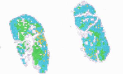
Image credit: The University of Queensland
News • Transforming medical diagnosis
Digital pathology: set to be a game changer in the medical industry
Patients will receive faster and more accurate pathology results following a decade-long research project that is set to transform medical diagnosis.
The University of Queensland (UQ) and Sullivan Nicolaides Pathology (SNP) have automated a microscope scanning and analysis system in Brisbane that has been tested, implemented and accredited ready for rollout around the world.
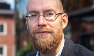
UQ Professor of AI Brian Lovell said the system significantly improved tests in terms of cost, quality and speed. “This digital pathology technology processes thousands of tests a day and has been accredited by the National Association of Testing Authorities (NATA),” Professor Lovell said. “At times the system can increase the productivity of pathologists and scientists by factors of 10 or more. The system also provides the ability to obtain second opinions via telepathology and dramatically improves record keeping and access of historical records, as the glass slides are no longer needed to be archived for years.”
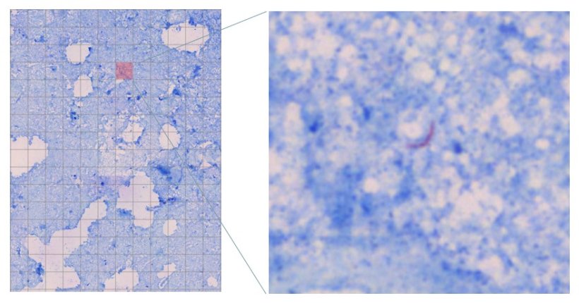
Image credit: The University of Queensland
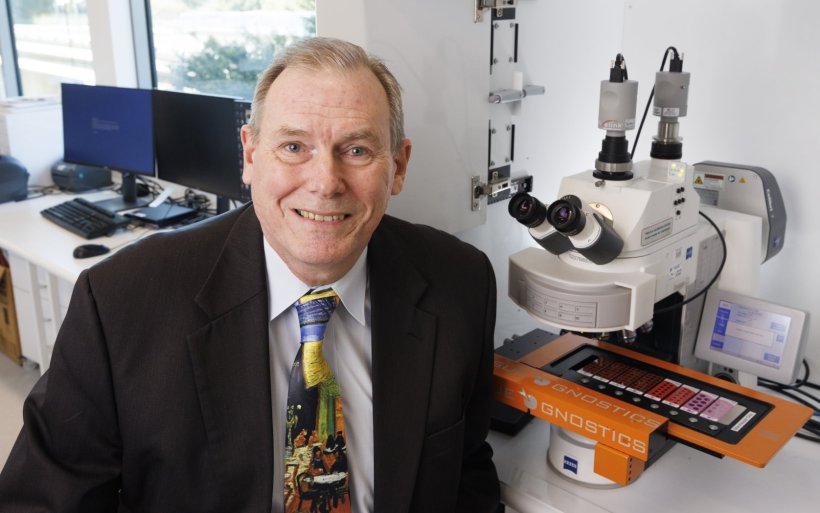
SNP Chief Executive Officer Dr Michael Harrison said the technology is a game changer in many areas of healthcare. “SNP laboratories in Brisbane are already using the system to improve the speed and accuracy of diagnoses,” Dr Harrison said. “Our scientists now use a digitised image often with associated AI instead of being tied to a microscope for many hours.”
Professor Lovell said there had previously been major problems with obtaining sharp, in-focus images with no human intervention. “Digital pathology images are often thousands of times larger than typical digital photos,” he said. “This had meant microscopy for diagnosing from tissue, blood and other specimen types was unable to be automated until now. Our active scanner knows what it is scanning and where it should scan, using image analysis and artificial intelligence. This greatly increases image quality and reduces file size.”
CEO of UQ commercialisation company UniQuest, Dr Dean Moss, said the technology demonstrated the benefits of industry collaboration with innovative researchers. “It’s exciting to see the advancement of a project that promises to have a transformative impact on better health outcomes,” Dr Moss said.
This research is supported by SNP, two Australian Research Council projects, and an Advance Queensland Fellowship from the Queensland Government. The technology won the Business and Industry Solution category at the recent Queensland iAwards, progressing to the national finals later this year.
Source: University of Queensland
21.07.2023