


News • Predictive phenotypes
Ovarian cancer: new insights into immune landscapes
Defining four different immunologic subtypes of recurrent ovarian cancers, researchers pave the way for more personalized treatment.


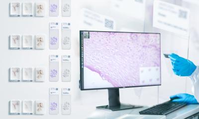
Step aside, glass slides: Digital pathology opens up a whole new world of possibilities in diagnosis, prognosis, and prediction of diseases, which is only furthered by the use of AI and Machine Learning. In our information channel, we have assembled some of the most exciting applications for digital pathology.
Defining four different immunologic subtypes of recurrent ovarian cancers, researchers pave the way for more personalized treatment.
Digital pathology software company Fujifilm Healthcare Europe and Ibex Medical Analytics, specializing in AI-powered cancer diagnostics, announce a formal partnership to support efficient and accurate cancer diagnosis.

North Bristol NHS Trust and Fujifilm Healthcare Europe continue their long-standing relationship, with the establishment of a reference site agreement for the company's new pathology solution.

The evolving role of AI tools in digital pathology was explored at an open discussion during the annual Digital Pathology and AI Congress in London with a high-level panel of practitioners looking at current and future technology options. The panel of pathologists, scientists and academics from Europe and the USA assessed the tools they currently use and are available to them, and those they…

Point-of-care diagnostics based on a combination of mobile-sized scanners and artificial intelligence (AI) are helping save the lives of women in low-resource settings. The AI technique is being applied in Kenya and Tanzania to deliver screening for cervical cancer – now the leading cause of cancer-related deaths in women in that region and a bigger cause of death than childbirth.

A new study measured how well breast cancer patients’ tumour ‘explants’ respond to chemotherapy or HER2 antibody therapy in the laboratory. This could help improve clinical outcomes.

Multiplex imaging can play a critical role in unravelling the tumour microenvironment. The potential and benefits of the emerging approach – a way to extract information from human tissue samples by visualising many more biomarkers than traditional microscopy – was highlighted in presentations during the 36th European Congress of Pathology in Florence, Italy. Speakers also discussed novel…

Artificial Intelligence (AI) remains a divisive topic within the discipline of pathology with a range of opinions over its current value and applicability in clinical settings. While most experts agree that the technology will not replace pathologists, it might still spell bad news for those who do not embrace AI in their daily practice. On the other hand, reservations persist about whether…

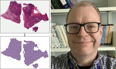
Cologne researchers have developed an AI-based digital pathology platform to enable extremely fast and accurate fully automated analysis of tissue sections from lung cancer patients.

New research shows that the AI large language model ChatGPT can be tailored to provide accurate responses to questions about digital pathology and compile detailed results.

Digital pathology can be used to great effect in pharmaceutical research: it can accelerate analyses, give deeper insights into cellular mechanisms, and enable better understanding of their role into clinical development. This potentially offers clearer predictions on how patients may respond to treatment and lead to personalized therapies.
Researchers work on the first prototype that applies AI to colorectal diagnosis. The prototype achieved a diagnostic acuity of 93.44% and a sensitivity of 99.7% in the detection of high-risk lesions.

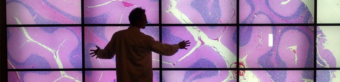
Researchers have unveiled a detailed understanding of immune responses in cancer, potentially paving the way for the development of new therapeutic strategies, the team hopes.

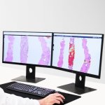
Digital pathology brings benefits for sample management and optimisation, lets pathologists work on samples remotely: The UK Government has now approved the use of the technique for cancer samples.

AI models are highly capable in analysing tissue samples – as long as conditions are lab-perfect. Add a little contamination, however, and diagnostic accuracy goes out the window, a new study shows.

Bringing digital pathology together with novel multiplexed staining techniques may answer key questions about complex diseases. Pathologist Lukas Marcelis, MD, PhD, believes such combinations of technology will have benefits for clinicians and patients and can help unravel some of the mysteries surrounding a range of conditions.

Centralized review of slides combined with telepathology has opened up the potential for a dramatic reduction in the waiting times for breast cancer patients to start their therapy. Jan Hudecek from the Netherlands Cancer Institute outlined his team’s framework for multi-centre clinical trials with centralized digital pathology review at the 9th Digital Pathology and Artificial Intelligence…

Experts have highlighted how precision pathology using Artificial Intelligence can provide an effective alternative to molecular diagnostics. This, say a team from the Karolinska Institutet (KI) in Stockholm, Sweden, can also offer multiple advantages within a clinical setting and support risk stratification.

With the introduction of digital pathology, the University of Queensland and Sullivan Nicolaides Pathology aim to provide significantly improved tests in terms of cost, quality and speed.