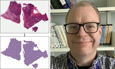
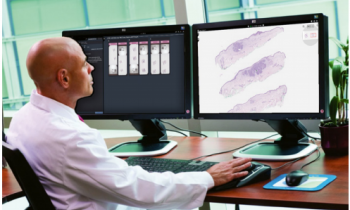
Image courtesy of Dr Alex Wright, University of Leeds
Article • Preparing for the future
Digital pathology dawns in developing countries
Pathologist Dr Talat Zehra reports from Pakistan

"Given the rapid transition towards digitisation, digital pathology is now unquestionably the future. However, some pathologists, particularly in underdeveloped countries, are still reluctant to accept its place in their labs. Among their many reasons, some feel that histopathology is a very complex and subjective field and artificial intelligence (AI) software cannot cope with all the issues. Conversely, pathologists are also scared that AI might replace them completely.
Last but not least, a large number of pathologists, particularly senior ones, or those who have not worked in the developed world, are not familiar with new modalities, so they are reluctant to adopt them. As for Pakistan, the bottleneck here is either the absence of pathology slide scanners, digital microscopes or manual whole slide image (WSI) software; if available they are mainly used for educational or research purposes only. The use of AI is almost negligible, indeed very few pathologists know about this novel entity.
Initially, I did some pilot projects to validate the results of AI software on previously diagnosed cases. For this I am highly thankful to Aiforia Technologies Oy, which gave me its demonstration version and training on AI software. Using this, I carried out some pilot projects on chorionic villi and malarial parasite detection and gained a good concordance of around 84%. We accomplished this project without a scanner or digital microscope.
Despite slow adoption in many institutions and countries, the digital slide image is slowly replacing the glass slide. Aided by AI-based image analysis software we can improve the much weaker and fragile healthcare delivery in developing countries. These carry most of the world’s endemic disease workload but, unfortunately, being less equipped with diagnostic techniques, the results are delayed diagnoses which, in turn, are associated with increased morbidity and mortality rates.
The digitisation of pathology resources opens the door for the use of the massive potential of AI in pathology, which can speed up more accurate and reliable diagnosis. Additionally, integration will help to ease large data storage and predict the future outcome of disease, as well as personalise care plans for each patient, and forecast oncology trends particular to specific geographies. In this way, pathologists could suggest proactive approaches to prevent disease progression. Thus AI-enabled digital pathology use will transform the horizon of pathologists’ traditional clinical practice, which has been unchanged for many decades.
The rise of the pandemic that penetrated the globe without discrimination, further enhanced the need for telepathology, which I and other pathologists belonging to my organisation use to send digital images to senior colleagues in hospitals in Karachi to avoid unnecessary travel and exposure. However, the bottleneck in most of our setup has been the lack of pathology slide scanners or digital microscopes through which WSI can be sent easily. So, the idea of ‘working from home’ through devices emerged, which resulted in new norms in the thought process of pathologists, even in Pakistan, and will leave its impact even after the pandemic completely resolves.
Many times, senior pathologists ask us to come to their location because digital images are not enough to give a reliable consultation, particularly in difficult cases
Talat Zehra
Additionally, an important and serious issue in digital pathology adoption is that a large number of technical, ethical and legal questions still need to be answered, particularly in developing countries with a weak monitoring system. A very important issue, which may not be a very big deal for developed countries, is the high cost of scanners and AI-based software – among the reasons for delayed adoption of low resource setups and countries.
That accounts for the delayed adoption of this novel technique in our area of the world. We sent the cases for second opinion in the form of digital images that we took through microscope cameras, which cannot produce a detailed image; so a second opinion is not always easy. Many times, senior pathologists ask us to come to their location because digital images are not enough to give a reliable consultation, particularly in difficult cases. So, due to the lack of scanners and digital microscopes, we have used digital images largely for research and educational purpose, not for routine clinical diagnosis.
The role of world leading organisations of slide scanners and of AI software could be of great importance. They could participate with pathologists and technical staff in developing countries, which are potential sources of big data, in terms of training, as well as to offer scanners, digital microscopes and software at a relatively affordable price, i.e. to put them within the budgets of low resource organisations.
Finally, I will conclude on the note that digital pathology and AI is in the forefront of modern pathology and its emerging use within healthcare is now being realised. The novel technology is at our doorstep and it is better to welcome and accept it, otherwise a large proportion of the world population will suffer.”
Profile:
Dr Talat Zehra MBBS FCPS is assistant professor at Jinnah Sindh Medical University (JSMU) and consultant Histopathologist at JSMU Diagnostic Lab, Karachi, Pakistan, She gained her Bachelor of Medicine, Bachelor of Surgery degree in 2007 from Dow Medical College, University of Karachi, Pakistan. She did her fellowship in Histopathology at the College of Physicians and Surgeons of Pakistan. Zehra’s field of interest is digital pathology and the use of artificial intelligence in tissue imaging. She has written a few write ups internationally to highlight the issues in delaying adoption of digital pathology techniques in developing world. Currently, she is also a member of the education committee of the Digital Pathology Association (DPA)
15.02.2021


