News • Assessment of tumour-infiltrating lymphocytes
Skin cancer: AI assistance helps pathologists identify melanoma
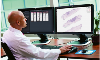
Pathologists' examinations of tissue samples from skin cancer tumours improved when they were assisted by an AI tool.

Image source: Karolinska Institutet; photo: Niklas Elmehed
The assessments became more consistent and patients' prognoses were described more accurately. This is shown by a study led by Karolinska Institutet, conducted in collaboration with researchers from Yale University, and published in the journal JAMA Network Open.
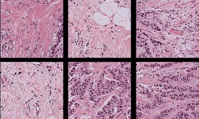
It is already known that tumour-infiltrating lymphocytes (TILs) are an important biomarker in several cancers, including malignant melanoma (skin cancer). TILs are immune cells found in or near the tumour, where they influence the body's response to the cancer. In malignant melanoma, the presence of TILs plays a role in both diagnosis and prognosis, with a high presence being favourable. An important part of pathologists' work in malignant melanoma is to estimate the amount of TILs. Researchers at Karolinska Institutet have now investigated how pathological assessments were affected by an AI tool trained to quantify TILs.
The study included 98 pathologists and researchers from other professions divided into two groups. One group consisted solely of experienced pathologists. They worked ‘as usual’, i.e. they looked at digital images of stained tissue sections and estimated the amount of TILs according to current guidelines. The second group included pathologists, but also researchers from other professions – all of whom had some experience in assessing pathological images. They also looked at the images ‘as usual,’ but were assisted by AI support that quantified the amount of TILs. Everyone assessed 60 tissue sections, all from patients with malignant melanoma. The study was retrospective, so the images showed tissue samples from patients whose diagnosis and treatment had already been determined.
Understanding the severity of a patient's disease based on tissue samples is important, among other things, for determining how aggressively it should be treated
Balazs Acs
The assessments made with AI support were superior to the others in several ways. Among other things, reproducibility was very high – the results were very similar regardless of who performed the review. This is important because assessments of TILs can currently vary depending on who performs them, which can compromise medical safety. The AI-supported assessments also provided a more accurate picture of the patients' disease prognoses – since the study was retrospective, there was a ‘correct answer’ to compare with. However, this outcome was unknown to those who assessed the images.
"Understanding the severity of a patient's disease based on tissue samples is important, among other things, for determining how aggressively it should be treated. We now have an AI-based tool that can quantify the TIL biomarker, which could help with treatment decisions in the future. However, more studies are needed before this AI tool can be used in clinical practice, but the results so far are promising and suggest that it could be a very useful tool in clinical pathology," says the study's last author, Balazs Acs, associate professor at the Department of Oncology-Pathology at Karolinska Institutet and a clinically active pathologist.
The research is funded by the Swedish Society for Medical Research and Region Stockholm, among others, as well as several grants from the US National Institutes of Health. See the study for any conflicts of interest.
Source: Karolinska Institutet
04.07.2025


