Article • Computational pathology
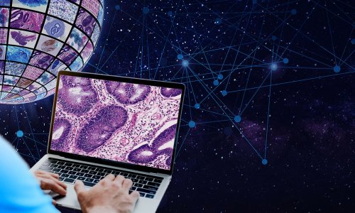
The tipping point for digital pathology
Digital pathology has been the next big thing for about a decade. Yet, today only a few pathology laboratories are fully equipped to digitise their workflow, mainly for legal or financial reasons.
In the USA, for example, the ‘struggle’ with the FDA has prevented large-scale introduction of whole slide images (WSI) in pathology for years. Nevertheless, it remains to be seen whether many pathology labs would invest the required (large) amount of money in a technique that still has to substantiate those high expectations.
Will WSI be the disruptive innovation, as some experts believe? I think it will, but only under the right conditions. Yes, it may streamline the laboratories’ diagnostic workflow, but only if fully and properly integrated with current workflows and information systems within the pathology department. WSI may be disruptive in the way we practice pathology at large, but only if we can create the right infrastructure to support networks of collaborating pathologists. Then it will be possible to practice ‘pathology in the cloud’ and instantly reach the right (subspecialised) pathologist for every difficult case. Disruptive as this organisation may be, perhaps the most important promise of digital pathology is the development of computational pathology algorithms for WSI. Once a computer can help a pathologist to interpret histopathological images, digital pathology will realise its full potential. Almost all research so far has been devoted to quantifying immunohistochemistry. The next step will be assessment of regular H&E stained sections. There are three (overlapping) areas in which computational pathology will have a major affect pathology diagnostics, with increasing level of impact and complexity.
- Computational pathology will increase efficiency of routine tasks. The first use cases for computational pathology will probably address tedious routine diagnostic tasks needing great accuracy, e.g. finding metastases in lymph node sections, or counting mitotic figures in breast cancer sections. Most pathologists are not fond of such tasks though they must be done well – they have high relevance for patient treatment planning. A computational pathology algorithm that can automatically detect metastases or count mitoses will alleviate these tasks and increase accuracy.
- Computational pathology can improve accuracy of tasks in which some grading is involved. Pathologists possess, at best, moderate reproducibility in such (semi-)quantitative tasks. A well-known example is Gleason scoring for prostate cancer. Computational pathology may offer a powerful alternative, by quantifying tissue changes that correspond with tumour grade in an accurate and reproducible manner.
- Computational pathology may yield relevant information for diagnosis and prognosis that the human eye and mind are unable to recognise or appreciate. Instead of using a computer to mimic a pathologist in, for instance, grading a tumour, we could also try to obtain relevant quantitative data directly from WSI. These ‘imaging biomarkers’ may drastically change the way we extract information from tissue sections. While promising, a significant amount of research and validation is needed before a patient benefits from this type of application.
Researchers will have to develop and validate algorithms before computational pathology will really take off. Deep learning, a modern pattern recognition technique, is extremely powerful in many different disciplines and for a wide variety of problems. We have recently shown (Litjens et al. Scientific Reports 2016) that deep learning is also particularly suited for analysing WSI. We found that deep learning yielded computational pathology systems that are close to being clinically useful.
The ‘Camelyon’ grand challenges
To get a broader view and establish the current state-of-the-art of computational pathology for a specific application, we organised the ‘Camelyon’ grand challenges (http://camelyon16.grand-challenge.org and http://camelyon17.grand-challenge.org) – a valuable instrument in medical image analysis, in which every researcher or research group is invited to develop an algorithm for a given problem. All participants solve the same problem, using the same set of data, and the challenge organisers evaluate all submissions in exactly the same manner. Therefore this is a great way to compare different approaches to problem solving.
In the Camelyon16 challenge, we offered participants a large number of full WSI of sentinel lymph node sections of breast cancer patients with exhaustive annotation of all metastases. We collected tissue sections in two different Dutch hospitals and scanned these on two different WSI scanners. Participants used these WSI to develop computational pathology algorithms. In the next stage of the challenge, participants ran their algorithms on a separate set of 130 sentinel lymph node WSI (this time without the ‘ground truth’ annotations) and we received their results for evaluation.
My strong conviction is that implementing computational pathology will tip the business case completely and even improve pathology diagnostics. But a significant amount of work is needed.
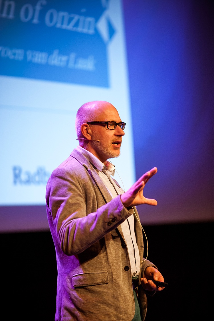
Jeroen van Laak
An important result is that many strong research groups worked on a very specific, clinically relevant application in histopathology, with a large set of fully annotated WSI, in a direct comparison. This definitely sped up developments, challenging researchers to spend significant time on a problem they would otherwise not tackle. A number of strong algorithms for this task resulted. Deep learning was applied in the top 10 algorithms, underlining the superiority of this technique for computational pathology. To our surprise, the best algorithms in Camelyon16 performed almost at the level of a pathologist participating in the challenge! We had expected computational pathology to be a powerful and promising development but hadn’t expected this level of performance at this stage. However, before we can start implementing computational pathology we have to validate these algorithms rigorously on a large number of cases.
My strong conviction is that implementing computational pathology will tip the business case completely and even improve pathology diagnostics. But a significant amount of work is needed. Development and validation of computational pathology algorithms mostly require the involvement of expert pathologists. However, the rewards are huge: a modern, accurate and reproducible assessment of human tissues facilitating the best treatment of every individual patient.
Profile
An associate professor and group leader at the pathology department in Radboud University Medical Centre (RUMC) in Nijmegen, The Netherlands, Jeroen van der Laak PhD Msc (computer science) leads a computational pathology research group. Internationally rated among the leaders in this field, the research team develops and validates deep learning algorithms to improve efficiency in pathology diagnostics. He has co-authored over 80 peer-reviewed publications, is a member of the Editorial Boards of Laboratory Investigation and of the Journal of Pathology Informatics and sessions organiser for the European Congress of Pathology and Pathology Visions. In 2016 and 2017 he coordinated the Camelyon grand challenges.
03.09.2017