


News • Body composition radiodensity
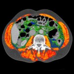
New CT-based marker to refine gastric cancer prognosis
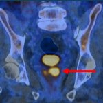
Measuring radiodensity of visceral fat and muscle from CT scans, researchers have identified a new biomarker that may help determine the prognosis for patients with gastric cancer.