Article • Machine learning advances diagnostics and prognostics
Computerized image analysis can predict cancer outcomes
The advent of digital pathology is offering a unique opportunity to develop computerized image analysis methods to diagnose disease and predict outcomes for cancer patients from histopathology tissue sections. Such advances can help predict risk of recurrence, disease aggressiveness and long-term survival, according to a leading expert in the field, Professor Anant Madabhushi from Case Western Reserve University in Ohio.
Report: Mark Nicholls
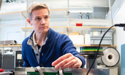
Speaking to online delegates at the 7th Digital Pathology and AI Congress, Professor Madabhushi – who is also Director of the Center for Computational Imaging and Personalized Diagnostics at Case School of Engineering - outlined how tools are being developed to advance this area of diagnosis, prognostics and prediction.

His team has developed a suite of image processing, computer vision and machine learning tools specifically designed to predict disease progression and response to therapy via the extraction and analysis of image-based histological biomarkers derived from digitized tissue biopsy specimens. They have already been applied in the context of several different disease domains including breast, lung, oropharyngeal, prostate, ovarian cancer, and endomyocardial biopsies. He suggested these tools would serve as an attractive and cost-effective alternative to molecular based assays, which attempt to perform the same predictions.
His presentation, “Computational Pathology as a Companion Diagnostic: Implications for Precision Medicine”, looked at the implications of such tools for precision medicine against a backdrop of cancer diagnosis and mortality in the United States. But with 600,000 deaths annually and 40% of the population receiving a cancer diagnosis, he noted: “There is some discrepancy between those two statistics. Some of the reasons why diagnostic incidence is high and mortality for the disease not in the same ball park is because we have become more aggressive about cancer detection, screening and imaging.”
Professor Madabhushi warned that overdiagnosis is harming patients – not only physically with the treatments and side effects of therapy, but also financially with a high proportion of cancer patients having to invest their life savings in their treatment. While finding disease earlier leads to a more favourable outcome, he suggested computerized image analysis could play an increasingly important role in the diagnosis of cancers and assessing the value of therapies.
Recommended article
News • RadClip
AI tool for MRI could transform prostate cancer surgery, treatment
Researchers at the Center for Computational Imaging and Personalized Diagnostics (CCIPD) at Case Western Reserve University have preliminarily validated an artificial intelligence (AI) tool to predict how likely the disease is to recur following surgical treatment for prostate cancer. The tool, called RadClip, uses AI algorithms to examine a variety of data, from MRI scans to molecular…
Immunotherapy is a game-changer for cancers such as melanoma and lung cancer, he said, but they are expensive and have a limited success rate of 20-25% and no clarity on which patients will benefit or respond to these therapies. He said that underlines the need for better diagnostic, prognostic and predictive tools that will identify the presence of disease, and predict disease outcome and progression, and predict the response to treatment. “AI and deep learning and machine learning can really aid the pathologist, but there is a real unmet clinical need for these tools which can benefit clinician, the person who is interfacing with the patient and having to make treatment and management decisions. This will support precision medicine by using prognostic and predictive tools for tailoring therapy for a given patient based off a specific risk profile.”
His group is working to understand the full value to be gained from the data gleaned from tissue biopsies and the unprecedented opportunity to use computational machine learning tools on the digitized slides. “This will not just identify presence or absence of disease,” he said, “but mine data for digital interrogation of data from perspective of mining digital biomarkers to identify features that can tell us about the risk of progression of disease, aggressiveness of disease, and how likely patients are going to respond to chemotherapy or immunotherapy.”
To illustrate this potential, he highlighted a series of studies from his group where AI and machine learning had been shown to suggest which patients would benefit most from which treatment. These included one focusing on disorder of collage fiber associated with risk of recurrence in Estrogen Receptor Positive (ER+) breast cancers in ECOG-ACRIN E2197 & TCGA. “This goes to show the value of these diagnostic tools to aid the clinician in figuring what the outcome is and how to treat these patients,” he said. “This is also true in early-stage disease.”
Computational analysis with routine imaging could help address questions in precision medicine, specifically prognosis and predicting response to therapy
Anant Madabhushi
In other examples, he outlined how a combination of computer extracted features of nuclear morphology, tubular formation and mitotic count predict disease free survival in ER+ breast cancer, and also how computer extracted images of nuclear shape, orientation disorder and texture from whole slide imaging are associated with disease free survival in ductal carcinoma in situ. He further detailed possibility of using similar approaches predicting post-surgical recurrence in prostate cancer, lung cancer, ovarian cancer, endometrial and cervical cancer.
In conclusion, Professor Madabhushi said: “Computational analysis with routine imaging could help address questions in precision medicine, specifically prognosis and predicting response to therapy.” The relatively low-cost aspect of computational diagnostic tools could also have global benefits, especially in low- and middle-income countries. “Also,” he added, “the importance of establishing the connection to the molecular underpinning of the features, moving away from an abstract representation, is going to be a really important driver for clinical adoption and clinical utility.”
Profile:
Anant Madabhushi is the Donnell Institute Professor of Biomedical Engineering, Case School of Engineering, Case Western Reserve University, Cleveland, Ohio, and also Director of the Center for Computational Imaging and Personalized Diagnostics. In the School of Medicine, he is Professor in the Departments of Pathology, RadiationOncology, Radiology, Urology and General Medical Sciences. His research interests include quantitative image analysis, radiomics, pathomics, cancer imaging, and digital pathology.
15.01.2021