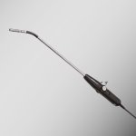
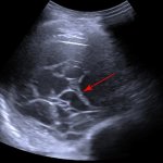
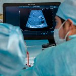
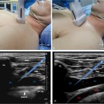
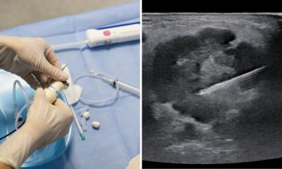
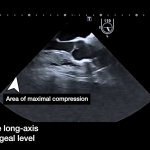
Photo:

Ultrasound

Ultrasound imaging is primarily known from prenatal care, but sonography is also used in many other places: it is just as indispensable for examining the thyroid and abdomen as it is in cardiology. Read all about application examples, new technology and more.

Photo

News • ‘E’ for Expertise

New AI-powered ultrasound device presented at SIRM 2024

At the SIRM Congress 2024 in Milan, Esaote presents its brand-new MyLab E80, an E-series ultrasound device designed for professionals.

Photo

News • Brain stimulation

Treating neurological diseases with 3D ultrasound

Researchers have developed a technology for targeted stimulation of the brain with ultrasound. This could be used to treat diseases and conditions such as epilepsy, Parkinson’s disease, depression,…

Photo

News • Artificial intelligence application

AI integration into cardiovascular ultrasound to improve diagnoses

Philips announced its latest AI-enabled cardiovascular ultrasound platform to help speed up cardiac ultrasound analysis with AI technology and reduce the burden on echocardiography labs.

Photo

News • High-resolution imaging

Ultrasound reveals vascular details with microbubbles

Microbubbles in a contrast agent allows for ultrasound imaging of the finest vascular details. This is the working principle of a technique called ultrasound localization microscopy (ULM).

Photo

News • Medical workflow assistance

AI and robotics support for ultrasound imaging

Autonomous robotic ultrasound systems could perform routine examinations and support doctors in the OR. New research shows that these systems can make everyday life easier for medical professionals.

Photo

Video • Transthoracic ultrasound localization microscopy

Super-resolution imaging of microscopic heart vessels

Researchers were able to produce sub-millimetre resolution images of cardiac micro-vessels. This non-invasive new technique could allow scientists to study the physiology of the heart in more detail.

Photo

Article • Point-of-care ultrasound in trauma

Returning e-FAST ‘to its roots’

Stagnation, under-use, unfulfilled potential: At the EUSEM congress in Barcelona, leading emergency physician Dr Joseph Osterwalder describes how e-FAST (Extended Focused Assessment with Sonography for Trauma) – a key point-of-care ultrasound technique for trauma – has changed over the last two decades, and not necessarily for the better.

Photo

Wearable monitoring device

Wireless ultrasound patch monitors vital signs in deep tissues

A team of researchers and scientists from the University of California, San Diego (UCSD) have developed a stick-on ultrasound patch, the first fully integrated wearable system for evaluating cardiovascular function while people walk, run, or even ride a bicycle. This Ultrasonic system-on-patch (USoP) is the result of several years of research conducted by nanoengineering department engineers.

Photo

Article • Portable imaging

Going mobile: advances in point-of-care ultrasound

Ultrasound technology now plays a vital role in clinical diagnosis and management. Significant advances in point-of-care ultrasound (POCUS) have made it a versatile tool for assessment, diagnosis, and follow-up across various fields. New developments continue to expand its applications, improving patient care and outcomes.

Photo

News • Imaging of focal breast complaints

Study confirms ultrasound as effective modality for diagnosing localized breast lumps

Ultrasound is an effective standalone diagnostic method in patients with focal breast complaints, according to a study published in Radiology, a journal of the Radiological Society of North America (RSNA). Focal breast complaints can refer to pain, lumps, nipple discharge or other symptoms and conditions confined to a specific area of the breast.

702 show more articles
Subscribe to Newsletter