


News • Hybrid deep learning approach
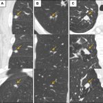
"Double vision" AI system to detect lung cancer from CT scans
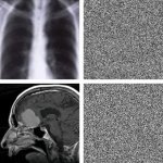
To improve lung cancer detection, researchers have developed a new AI system that employs a dual approach to analyse CT scans – seeing both detail and context at the same time.