Microscopy
Digital pathology: a new diagnostic technology
Histopathologists play key roles in diagnosing disease entities and determining biomarkers related to the prognosis and response to specific therapy of malignant tumours.
Report: Bela Molnar

Histopathology is still firmly based on cell and tissue morphology supplemented with in situ molecular information and these together can be studied using an optical microscope.
Digital microscopy creates a digital representation of the whole microscopic slides at decent quality, which can be dynamically viewed, navigated and magnified via a mouse and computer monitor, and shared though computer networks without spatial and temporal limitations. Digital slides can be integrated into the hospital information system (HIS) and accessed through intra- or internet for teaching, primary diagnosis, teleconsultation and quality assurance.
Discrete pixels of calibrated qualities allow automated image analysis and signal quantification for drawing unbiased conclusions in diagnostic and research applications. Therefore, utilisation of the full power of computer technology to access multiple functions and the internet grants digital microscopy great potential to upgrade the efficiency of pathology workflow and pathologists. By resolving critical issues, including standardisation of data formats, secure and fast internet communication and medico-legal aspects, digital microscopy is expected to play a revolutionary role in future histopathology.
Digital slides created by slide scanners
Digital microscopy creates large digital files representing all crucial details of stained tissue sections with decent resolution and high colour fidelity achieved using automated focusing and white balance. Digital slides are made up of giant arrays of rectangular pixels organised along x-y coordinates, each of which is characterised by size, colour and intensity values.
Produced either by area or line scanning, digital slides are built up as pyramids of microscopic image series where low power views are generated by compressing the original sharp and optimally lit images (Fig. 1A). Scanning through several focal levels within the usual 3-8μm sample thickness offers access to the z dimension used for emulating fine focusing of the optical microscope. Fig. 1 A) The image series by compression allows viewing of the digital slide at arbitrary magnifications (e.g. x15 and x10). Scanning at different focal levels within the sample thickness (~3-8μm) offers access to fine details in the z-dimension.
Using x10 lens offers high field of view (FOV) size and focal depth, while requiring small storage space. A x20 objective allows double the optical resolution than that of x10, but at the expense of revealing smaller FOV and focal depth, while increasing the storage need. x40 objective does not offer significant improvement in optical resolution compared to x20 (NA=0.9 vs.0.8) despite needing large storage space and long scanning time.
Unique features of digital slides
Seeing slides on a monitor, with easy access to a computer’s multi-functionality, is far more ergonomic than peering though an ocular lens of an optical microscope. Even pathologists with high affection for conventional microscopy respect digital microscopy benefits if they practise enough. The computer-generated image pyramid format of digital slides allows in-focus navigation through continuously changing magnifications, without changing objectives, or realigning the focus or lighting conditions. Digital magnifications beyond that used for scanning still reveal fine microscopic details hidden in the original magnification.
Slides can be tilted arbitrary for proper orientation and preview images of the whole slide are available simultaneously on the monitor where navigation history of high power analysis can also be traced (Fig. 3 A). Digital slide viewer interfaces utilise the whole computer monitor where preview images and navigation history (left side) of high power analysis can also be traced. Calibrated pixels allow straight measurements of object distance, perimeter or area highlighted by permanent annotations. The monitor can be shared for several digital slides for comparative studies, as shown by the same area of serial slides stained for H&E, the proliferation marker Ki67 (brown; middle) and the gap junction connexin-43 (red immunofluorescence) combined with the Ki67 protein (green; right), respectively in oral epithelial hyperplasia. (Fig. 3B)
Permanent annotations and text put on digital slides, straight measurements of object distance, perimeter or area and prompt still-image archiving at publication quality all support the pathology workflow. Several digital slides can be opened side-by-side on the monitor for comparative analysis of serial slides of a sample stained for different biomarkers. Even samples of immunohistochemistry and FISH (fluorescence in situ hybridisation) can be opened, linked and navigated alongside, which normally need consecutive steps or even separate microscopes.
Pixels making up the images have calibrated dimension, colour and intensity, features that can be used for colour separation-based automated quantification and measurements of image-objects. Furthermore, pattern recognition of morphological and functional units within the tissue, such as glands, or hyperplastic or abnormally arranged epithelial nests, can be automatically made based on shape, size and texture identification.
Serial digital slides can also be assembled into a 3-D structure for reconstructing tissue architecture, e.g. to study tumour invasion or re-orient colorectal biopsies. Digital data, including whole digital slides with annotations and measurements, can be integrated into digital databases and shared though intra- or internet with unlimited partners, even simultaneously. The freedom to access digital slide archives for re-review, and the logistics of slide storage and sorting are simple tasks managed through a computer.
All these, of course, need advanced IT, including high speed computers, massive storage capacity, safe data handling using backup storage, and wide-range internet access and dedicated software tools with a user-friendly graphic interface.
Molecular pathology visualisation
Fluorescence microscopy detects fluorophores used to label molecules in cells and tissues with the techniques of molecular morphology. Fluorophores are activated at UV or visible wavelength to emit light of lower frequency, usually in blue, green or red, which can be collected through emission filters in a dark background.
Samples targeted with fluorescing labels, particularly genomic FISH, must be studied within a short time-frame to avoid false negative results due to rapid signal fading (fading artefacts).
In addition, small signals of a few hundred nm size, such as those of gene and chromosome probes in FISH, or those of its chromogenic version (CISH), are randomly placed throughout the whole 3-8 μm thickness of tissue sections, or cells, and thus some remain hidden from conventional single focus photography. These signals can only be revealed with confidence by scanning several focal planes through the sample (z-stacking, or extended focus) for proper quantification of gene/chromosome gain, such as that of HER2 on chromosome17 (CEP17) in breast cancer (Fig. 4A-F); or for the fine spatial localisation of signals proving gene-translocations, such as that of t (9;22) resulting in the BCR-ABL gene fusion in a case of chronic myeloid leukaemia. Gene abnormalities may determine a specific diagnosis and the concomitant treatment options in an increasing number of malignant tumours.
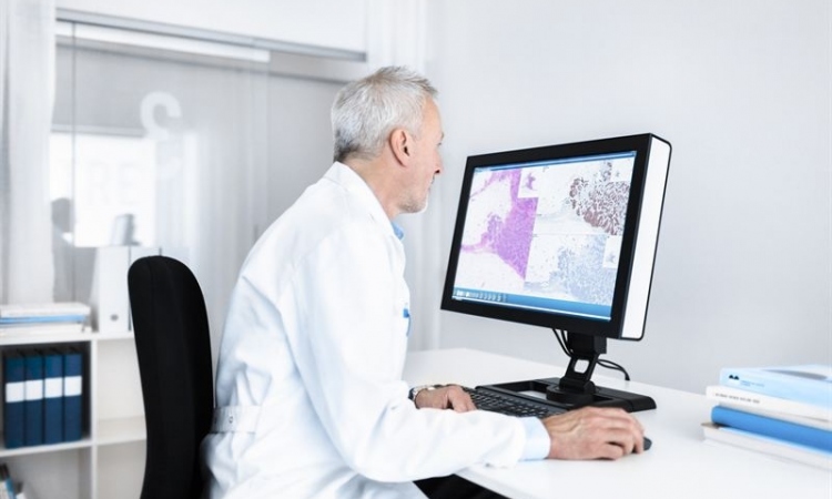
Digital pathology within routine sign out processes
Once the digital slide scanners digitise the hematoxilin eosin, surgical pathology specimen, and then immunohistochemical second round and FISH third round slides are also available in the slide holder/server solution, a pathologist can evaluate them from the laboratory’s digital workplace. (Fig. 5) In addition to slide viewing, the software supports quantification of identified alterations. The digital report can be signed out with detailed image and quantitative data.
PROFILE:
Bela Molnar MD DSc is CEO of 3DHISTECH Ltd, which has developed high-performance hardware and software products for digital pathology since 1996. A medical graduate from Semmeweis University, Budapest, and the Hungarian Academy of Sciences, he gained qualifications as an internist and gastroenterologist, followed by a post doctoral 2-year fellowship with Boehringer Ingelheim GmbH. Scientific/industrial cooperation arose with Roche Diagnostics, Epigenomics Inc (Seattle/Berlin), and Carl Zeiss MicroImaging. Today, Molnar’s main research areas include biomarkers of colorectal cancer development, molecular biology applications, virtual microscopy, and quantitative image analysis, and his publications, memberships of scientific bodies and professional awards are numerous.
23.05.2016