Image source: Philips
Article • Need for modernisation
Digital pathology: Luxury or necessity?
The anatomical pathologist faces a crisis. Public and private labs suffer increasing caseloads, whilst pathologist numbers diminish for various reasons, including greater cancer prevalence associated with aging populations as well as improved cancer screening programs.
Precision medicine typically involves more genetic testing and extensive use of immunohistochemistry to classify cancer and assess prognostic and predictive biomarkers. In clinical practice, a notable number of pathologists are nearing retirement, yet today’s diagnostic pathology training of young doctors is limited.
Alternative: the optical microscope
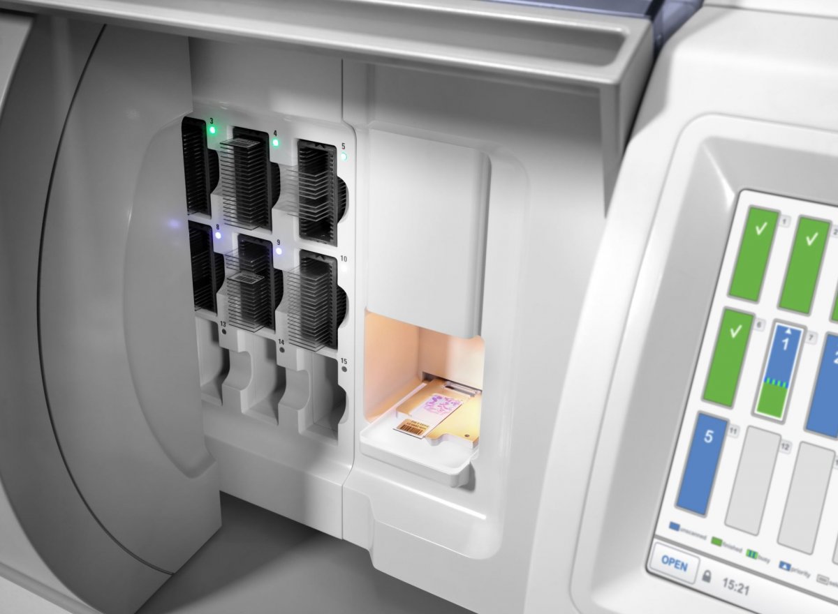
Image source: Philips
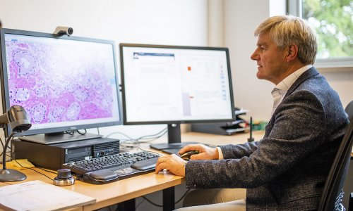
Recently, digitisation and digital pathology have become widely accepted due to advances in technology and regulations. In essence, in digital pathology a scanner produces a digital copy of the traditional glass slide, to be stored in a local or cloud-based server and viewed anywhere with a computer and internet connection. Current scanners provide high fidelity digital images thanks to high magnification (40x). Additionally, high throughput scanners, with a capacity of up to a thousand slides, are capable of fast, reliable operation with little human intervention, making possible unsupervised overnight operation and samples ready for review by pathologists and trainees in early morning.
Licensed but a slow uptake
In 2017, the USA’s Food and Drug Administration (FDA) warranted the first license for the use of digital pathology for primary diagnosis in that country. Permission for in vitro diagnosis came sooner in Europe. Despite this, adoption of digital pathology has been slow for several possible reasons. A commonly mentioned barrier is pathologists’ concerns, such as digital images inferiority, or a slowdown in the sign-out process.
Decades ago, when digitisation entered radiology to produce digital images, film was no longer needed. The savings eased an initial reluctance among some groups, but not in pathology since elaboration of the glass slide is still required. This, coupled with concerns over increased turnaround times due to the added step of scanning, as well as the substantial initial investment needed to fully digitise a lab, help explain the slow rate of path digitisation.
What’s in it for pathologists?
Despite concerns, today’s digitisation brings multiple advantages to pathologists and patients. Early digital pathology adopters report many benefits, including efficiency gains and better case allocation across digital pathology networks. The availability of a digital histology slide enables distant viewing and remote reporting, particularly appealing given demands imposed by COVID-19. Multiple users can access digital images, regardless of location, enabling second opinions and consultations. Shipping and storing glass slides on site is obviated. Digital rather than analogue archives are more manageable; prior cases are available quicker for comparative review.
Early users report overall efficiency includes shorter reading times during diagnostic sign-out sessions, improved overall lab efficiency in the range of 20%, or more, after complete digital diagnosis, compared with analogue workflow. In addition, the improved intra-laboratory logistics (the technician time required to sort the hundreds of slides prepared every day in the typical lab, distributing them to the responsible pathologist, collating them for tumour boards, and the added ease to manage consultations, etc.) may add to the savings and lower the financial barriers for adoption.
AI in pathology – friend or foe?
Artificial intelligence (AI) tools, including deep learning algorithms, can be applied to a digital histology image to facilitate computer-assisted diagnosis, which forms the basis of computational pathology. The application of AI to pathology bears promise, including increased accuracy in immunomarker quantification, better sample screening and pathologist efficiency, with reduced mis-diagnoses.
The discovery of new morphological predictors of disease outcome, invisible to the human eye but apparent to a computer, also suggests new diagnostic possibilities. The current capability to manage and analyse huge pathology image data, combined with more advanced techniques to simultaneously detect multiple molecules or antigens in situ, fosters a novel field of research. However, a prerequisite for computational pathology to fulfil its promises and become mainstream, is the general adoption of digital pathology – because the development and training of AI algorithms requires vast amounts of carefully annotated digital data. Additionally, once algorithms have been developed, they can only be applied in clinical routine use in labs that have successfully embraced digitization: by logic, AI tools cannot be applied to analogue glass slides. Some medical professionals fear that machines could work more rapidly and accurately than a human. However, the whole point of computer assisted diagnosis and computational pathology is to help pathologists.
In monetary terms
Digital pathology efficiency gains can be translated into monetary terms. It is important to estimate how long before an initial investment in lab digitisation show a return. A model proposed by the University of Leeds, in the UK, estimates that efficiency gains of between 10-15% result in an amortisation of the initial investment after 1-2 years. Based on this model, efficiency gains of 20%, as reported by some users, would point towards an early amortisation of the initial cost outlay.
How staff requirements change due to enhanced productivity in a fully digital lab needs analysis. Where pathologists are retiring and cannot be replaced, early digital pathology adopters report that, due to greater efficiency, a reduced pool of pathologists can absorb increasing caseloads. The differences in full-time equivalents (FTEs) of pathologists’ time to process the same caseload, when comparing those on the microscope, versus that same group of pathologists with better efficiency after digital transformation, show that the personnel savings from fewer FTEs needed are sufficient to amortise the initial investment after 2-3 years. Additionally, these FTEs savings, over five years – the typical life of a digital pathology system – do result in an additional lab profit over the fourth and fifth year, before additional expenditure is needed to renovate obsolete elements in the system. Therefore, the investment made efficiency gains that translate into a repayment of the initial amount around the second year and additional monetary gains for a further 2-3 years.
Can I work remotely?
A pillar of digital pathology is reliance on the creation of a digital twin of the glass slide, in a file accessed by anyone with the appropriate credentials from anywhere with an internet connection and a PC. The images are typically stored in a server, usually integrated within the main hospital infrastructure. From here, the digital slides can be accessed remotely via a virtual private network (VPN), that creates a secure access to the hospital IT system. The data transmission rate necessary for satisfactory viewing – a bandwidth of 300 megabits per second (Mbps) – is sufficient to ensure optimal performance when loading, panning and zooming the image. Currently, the typical optic fibre bandwidth for domestic use offers three times that speed.
Other important elements are the PC and monitor needed to view digital images. In 2017, the FDA warranted permission to the first digital pathology system for primary diagnostic use in the USA. Part of the system is a PC with an Intel Xeon CPU E5-1620 v3 at 3.50-GHz processor, 16 GB RAM, and an NVIDIA Quadro K4200 graphic card. These hardware elements, as usually with IT components, cost significantly less now. The FDA monitor approved for primary diagnosis is medical grade LED with a resolution of 1920x1200 pixels and a self-calibration mechanism. However, the emergency declared due to COVID-19 has resulted in a temporary relaxation of regulations pertaining domestic reporting, and currently user discretion is recommended when using domestic PC and monitors for home reporting.
In short, the IT infrastructure needed for remote diagnosis is not unduly demanding and can reasonably be met even by domestic users, which makes home reporting possible – particularly interesting in terms of social distancing rules due to the COVID-19 pandemic.

Profiles:
Falko Fend MD is Professor of Pathology and Chairman of the Institute of Pathology and Neuropathology and Reference Centre for Haematopathology at the Eberhard-Karls University in Tübingen, Germany. Following postgrad training in the Pathology and Internal Medicine Departments at Innsbruck University, in 1991 he became its staff pathologist. From 1997-1999, he was a research fellow at the Laboratory at the National Cancer Institute, NIH, Bethesda from 1997-1999, when he became Associate Professor of Pathology at the Institute of Pathology of the Technical University Munich, and later took on his current position at the University of Tübingen. His research focuses on the pathology and molecular genetics of malignant lymphomas and innovative molecular pathology. Fend is also a member of the executive boards of the German Society of Pathology, the European Association of Haematopathology and served as Chairman of the European Bone Marrow Working Group.

Dr Juan Antonio Retamero is an anatomical pathologist with extensive experience in the implementation and use of digital pathology for routine diagnosis. He played a key role in the adoption of digital pathology in a pioneering group of Spanish hospitals in 2016. He has shared those experiences internationally and supported many centres in adoption digital pathology adoption globally, including labs in the USA, Europe and Asia. He is a regular speaker at digital and computational events and an ardent advocate for modernisation of the profession. Retamero is also Consultant for Philips Digital and Computational Pathology.
17.09.2020