Article • Next-generation pathology
Multiplexed staining techniques in the fight against complex diseases
Bringing digital pathology together with novel multiplexed staining techniques may answer key questions about complex diseases. Pathologist Lukas Marcelis, MD, PhD, believes such combinations of technology will have benefits for clinicians and patients and can help unravel some of the mysteries surrounding a range of conditions.
Report: Mark Nicholls
In one example, he believes it could explain why younger patients with Epstein-Barr Virus-positive diffuse large B-cell lymphoma NOS (EBV+ DLBCL, NOC) generally fare better than older people. Marcelis discusses these technological advances in his presentation to the 35th European Congress of Pathology in Dublin entitled “Next-generation pathology using multiplexed immunohistochemistry in haematological malignancy.”
MILAN gives better context
A medical physician and trainee pathologist at University Hospitals Leuven in Belgium, having completed his PhD in EBV-driven lymphomas, he is continuing research into new multiplexed stain technologies. Speaking ahead of his presentation, he believes next-generation pathology using digital images analysis will offer significant benefits.
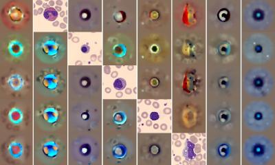
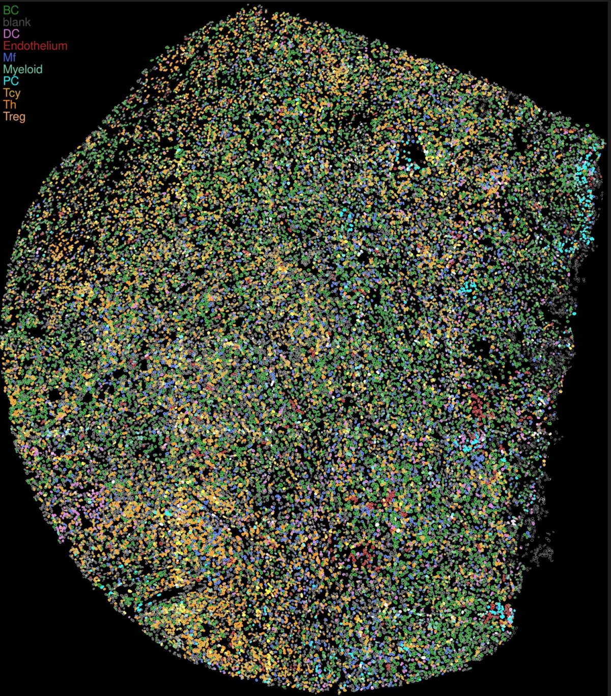
In a transition that sees pathologists no longer reading slides with their eyes but using computer digital image analysis, he points to the importance of correcting artefacts, and good quality control. ‘One advantage is that you can identify very specific immune cell subtypes, with spatial context,’ he added. With multiplexed immunohistochemistry allowing for significantly more markers on a slide (each cell can have 50+ markers), he has embraced the MILAN (multiple iterative labelling through antibody neodeposition) technique which uses immunofluorescent markers.
Potential in EBV-driven lymphomas
In haematological malignancy, he notes this is of particular interest in his research area of EBV-driven lymphomas, which can arise in immune-compromised patients, such as those receiving immune suppression to prevent organ rejection post-transplantation. He said: ‘One question is why some develop EBV-driven lymphoproliferative disorders (EBV-driven LPD) and others do not, and why sometimes if we reduce immunosuppression the disease disappears and sometimes not.
‘In EBV-driven LPD there is a lot of interaction between immune cells, the virus and the malignancy itself which requires a multitude of antibody stains to adequately characterize.’ The technique may help in predicting why in some, mainly younger, patients the immune system can control the condition better than in older people, for example.
Introduction into clinical practice almost within reach
A possible advantage for patients, he added, is that the microenvironment can be more fully characterized. This is currently highly interesting in research settings but while there is still much work to be done, he believes there is potential for this to be implemented into future clinical practice. Questions, however, remain around quality control, guidelines agreement and image quality.
For example, he stressed the importance of the machine adequately recognising individual cells and avoiding over-segmenting and splitting up one cell into different parts, or under-segmenting and grouping cells together. ‘Many different methods for cell segmentation exist, but for clinical practise a consensus on “gold standards” would be needed,’ he said. ‘But there is definitely a future in this digital image analysis using high-multiplexed stains because it can give a lot of information that cannot be obtained with classic immunochemistry.’
New answers (that will lead to new questions)
Dr Marcelis suggests there are benefits on a therapeutic and diagnostic level, such as in helping identify immune microenvironment “state” of the patient to predict the behaviour of EBV-driven LPDs or potential therapeutic options. ‘Next-generation pathology and digital image analysis and multiplex will allow to identify complex immune cell types in a spatial context and do neighbourhood analysis, all things not possible with traditional immunochemistry or flow cytometry.’ The big step is looking at a combination of 50-plus markers digitally on a single slide rather than through a microscope and examining and interpreting the slide differently. ‘Many questions will need to be answered,’ he said, ‘since these techniques often identify new immune cell clusters where we do not always know enough to name these cell types and if they are genuine or an artefact. But the future is that we are going to work more and more digitally and will have to use these techniques since they will have advantages for patients,’ he concluded.
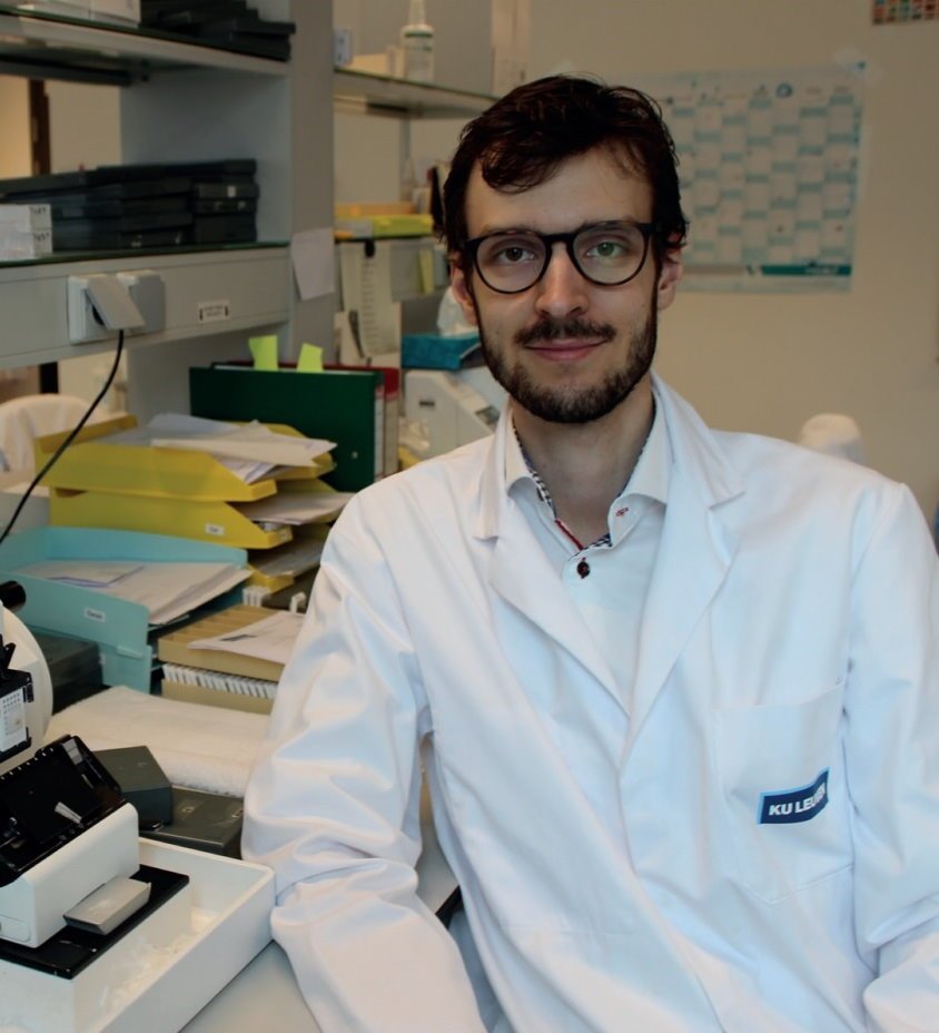
Profile:
Lukas Marcelis, MD, PhD, is a pathologist from the Department of Pathology at University Hospitals, Leuven, Belgium, having completed his medical training at the University of Leuven and obtained his PhD in the field of biomedical sciences on Epstein-Barr Virus-driven lymphoma. His research focus is on multiplexed stain technologies in haematological malignancy. A winner of the international David Y Mason award as a promising young researcher in the field of hematopathology, he is a co-founding member and secretary of the Young European Association for Haematopathology (Young EA4HP).
12.01.2024