
Microscopy

Photo

News • Stain-Free Histology

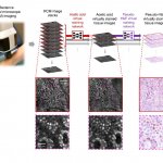
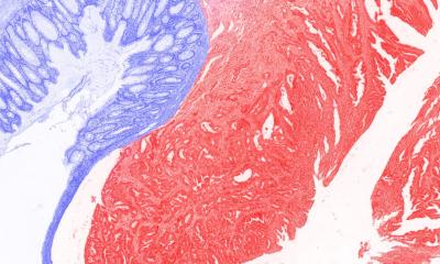
Nanophotonic slides add color to tissue samples

Researchers have developed a method to enhance the cellular features in pathology slides based on “structural color” – the same effect that gives peacock feathers their unique sheen.

Photo

News • Fluorescence lifetime imaging microscopy

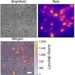
New technique could aid targeted lung cancer treatment

Detecting genetic mutations associated with lung cancer via laboratory tests can be expensive and time-consuming, and use up valuable tissue from small biopsy samples. A new approach aims to fix this.

Photo

News • Microscopy

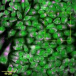
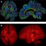
New microscopy method maps lipids without labels

A new microscopy method reads lipids' natural spectral fingerprints using mid-infrared light and ultrasound – no fluorescent labels needed, less stress for living cells.

Photo

News • Cryo-electron tomography

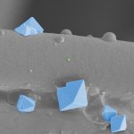
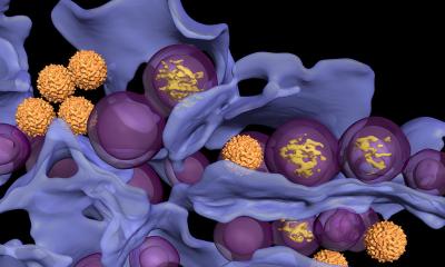
3D microscopy reveals how tick-borne virus replicates

Tick‑borne encephalitis is one of the most dangerous viral diseases spread in Europe. Researchers have discovered how the viruses replicate inside cells, paving the way for future treatments.

Photo

News • Fourier lightfield technology

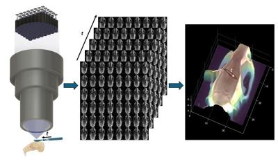
48-camera microscope offers precise 3D imaging during surgery

Using 48 cameras simultaneously to provide better insights during live surgery: A new kind of surgical microscope is designed to meet the need for better 3D imaging.

Photo

News • High-resolution cell scans

AI spots cancer and viral infections at nanoscale precision

Researchers have developed an artificial intelligence which can differentiate cancer cells from normal cells, as well as detect the very early stages of viral infection inside cells.

Photo

Article • Mini-microscopes

Are disposable robots conquering the labs of the future?

The team at the Institute of Medical Device Technology at the University of Stuttgart, Germany, is developing methods to produce top-quality medical devices at affordable prices. Professor Dr Peter Pott, the director of the institute, turns to 3D printers to successfully realize his vision of “high end at low cost”.

Photo

Article • Super-resolution miscroscopy

PEAR: setting nano-imaging in motion

Ever since the Abbe diffraction limit of conventional microscopy has been surpassed, super-resolution techniques have been diving ever deeper into the most miniscule details of molecular structures. We spoke with Prof. Dominic Zerulla, whose company PEARlabs is developing an imaging technique that sets out to push the boundaries once more – by looking at in-vivo nano-scale processes in motion.

96 show more articles
Subscribe to Newsletter