Automated DIA
Use the digital image analysis in Breast Cancer
In a study of 436 breast cancer cases with 28 years of survival data histo-pathologists from Karolinska Institute, Stockholm, Sweden shows that protein tissue-based biomarker data are at least as good as gene expression assays.
When summarizing the study from Karolinska Institute, Stockholm, Sweden, manual assessment of the biomarkers ER, PR, HER2, and Ki67, with an emphasis on the latter, is in most aspects an inferior alternative to Digital Image Analysis (DIA). When comparing sensitivity and specificity to PAM50 subtype classification and prognostic power DIA outperforms manual scoring. Being able to reduce time to diagnosis as well as the overall cost for breast cancer testing are good news. What makes it even more convincing is that all subtypes including the Luminal B, which is widely considered being the most challenging distinction in surrogate sub-classification, produced a slightly better concordance and Cohen’s agreement.
Digital Image Analysis (DIA) a viable and competitive, if not superior, alternative for gene expression testing in breast cancer
The cost of combining automated DIA with biomarker analysis is a fraction of the cost of a gene expression test, which is time consuming to conduct and often has to be done outside the histopathology lab. Combining biomarker analysis with DIA creates value for all parties involved: patients, pathologists and payers. For patients it is to receive a faster treatment, for pathologists an opportunity to reduce time consumption and allocate precious resources to more qualified tasks and for payers a much more cost efficient diagnostic method.
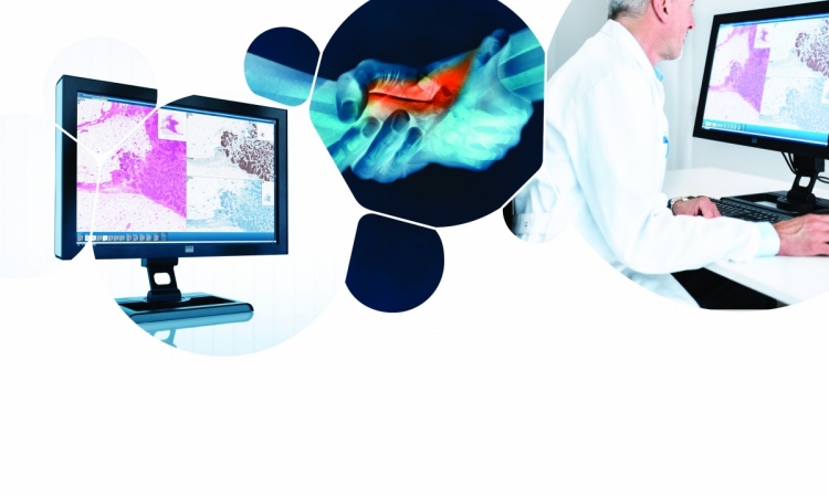
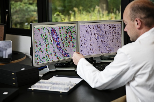
An operator of the Visiopharm DIA system has the option to manually evaluate the digitized slide image and perform the diagnosis based on this, but DIA also allows the computer to conduct an automated analysis avoiding subjective assessments of biomarker positivity. This implicates that an approach like DIA to count full tumor cross-sections or to make automated ‘hot spot’ analysis could free up time for histopathologists, biomedical scientists or other laboratory personnel.
“The desire for faster and more cost-effective solutions for the histopathology laboratories need no longer be a dream. In complex heterogeneous tumors DIA in combination with immunohistochemistry has shown to be a viable competitive option for laboratories lacking in-house gene expression testing,” says Helle Fisker, CMO at Visiopharm.
The DIA system used in the study was the Visiopharm DIA software VIS 4.6.3
23.05.2016
- economy (1049)
- histology (25)
- imaging (1676)
- IT (896)
- laboratory (1130)
- management (229)
- pathology (304)
- studies (1186)