
News • Coronary plaque vulnerability
Using radiomics to predict heart attacks
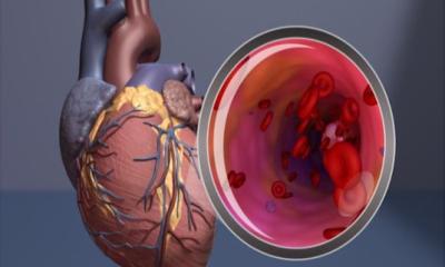
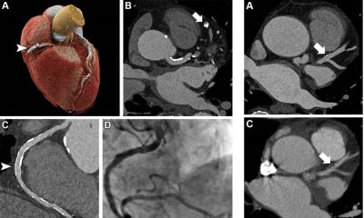
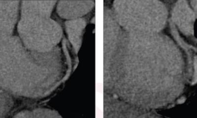
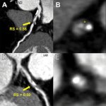
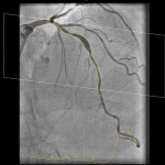
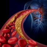
Researchers developed a radiomics model that uses information from coronary CT angiography images to assess coronary plaque vulnerability, a common cause of heart attacks.


Researchers developed a radiomics model that uses information from coronary CT angiography images to assess coronary plaque vulnerability, a common cause of heart attacks.
Photon-counting detector CT reduces the amount of contrast needed for CT angiography (CTA) while maintaining image quality, according to a new study published in Radiology: Cardiothoracic Imaging.
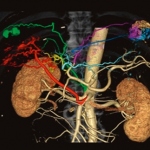
The system could enable significant advances for the 40,000 pediatric congenital heart disease patients born each year.

When a patient suffers a stroke, speed in treatment can mean the difference between successful recovery, permanent disability, or death. For Christopher Hess, success in stroke diagnosis is a question of workflow and efficient care delivery.
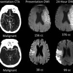
A commonly available imaging technique can identify stroke patients most likely to benefit from a procedure to restore blood flow, a potential breakthrough in care that could save thousands of lives every year

An AI-led device to assess coronary CT angiographs has been designed to assess cardiac plaque that may lead to myocardial infarction (MI). In his presentation ‘Vascular inflammation and cardiovascular risk assessment using coronary CT angiography’ (CCTA), Charambalos Antoniades, Professor of Cardiovascular Medicine at the University of Oxford, presented the research team’s findings during…
With interventional procedures becoming more and more complex the demands on the interventionalists are also increasing. Endovascular simulators allow practical angiography training. In December 2020, the University Hospital Essen, Germany, was the first European facility to install Mentice’s VIST G7+. Professor Dr Jens Theysohn, senior physician at the Institute of Diagnostic and…

Robocath announces the success of its first two coronary angioplasties performed with assistance from its R-One robotic platform.

Earlier this July, 75 Medical Physics Experts gathered in Prague to attend the EFOMP and COCIR Summer School “State of the art & new trends of angiographic equipment: Image quality, Patient and Staff dosimetry”, endorsed by the European Society of Radiology (ESR). The Summer School was organised in collaboration with the Czech Association of Medical Physicists and the Department of…

Siemens Healthineers and Mentice AB announced the collaboration to fully integrate Mentice’s VIST Virtual Patient into the Artis icono angiography system from Siemens Healthineers. The VIST Virtual Patient thus becomes a fully integrated simulation solution for the angio-suite. The global partnership between the two companies will allow interventional radiologists, neuroradiologists, and…
Canon Medical Systems Europe B.V. introduces a new angiography configuration featuring its Alphenix Sky+ C-arm and Hybrid Catheterization Tilt/Cradle Table for interventional procedures with its Aquilion One Genesis CT system. The new pairing, called the Alphenix 4D CT, allows clinicians to efficiently plan, treat and verify in a single clinical setting. “The all new Alphenix 4D CT was designed…

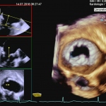
Intracardiac echocardiography (ICE) is an increasingly important guiding tool for structural heart disease interventions – without general anaesthesia. José Ribeiro, who works in the thorax and circulation unit at Gaia Hospital Centre, Portugal, who has worked with this technology for the past two years, explained its benefits and limitations.
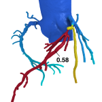
A new software to calculate the pressure drop and vFFR value (vessel Fractional Flow Reserve) in the coronary artery non-invasively was presented to interventional cardiovascular experts at EuroPCR 2018 in Paris. The software called CAAS vFFR (Cardiovascular Angiographic Analysis Systems for vessel Fractional Flow Reserve) was developed by Pie Medical Imaging (Esaote Group). The vFFR calculation…
Celebrating 40 years of PCI, cardiologists fret over their future with big data, machine learning and robots.

Coronary angioplasty is arguably the most revolutionary breakthrough in the history of cardiology. While the technique is today performed on millions of patients worldwide, its origins can be traced back to Zurich, Switzerland, in the late 1970s.
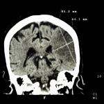
Stroke patients will first undergo a CT scan as they enter the hospital. Before any further imaging scan is carried out, the medical team must decide whether they need to intervene intra or extra cranially. ‘Imaging enables you to see which pathology you are dealing with and helps you select patients for either recanalisation or revascularisation or, in some cases, occlusion by embolisation,’…
One of the first facilities to purchase a complete set of the 3-D TEE transducer, including the equipment, was the Department of Cardiology and Angiology at University Hospital Magdeburg, as Thomas Groscheck, specialist physician for internal medicine at the echocardiography lab explains. Since July 2015 he has worked with the new Siemens transducer – and is enthusiastic.
Coronary interventions often rely more on art than science as the decision to treat a patient tends to be based on what clinicians can see, a subjective interpretation of cardiac imaging. Two new techniques have emerged for cardiovascular diagnostics that are enabling software to help surgeons and cardiologists measure, and thereby better manage cardiac disease.
Did we get all of the tumor during a cone beam CT? Can the patient hold his breath for several seconds during a CBCT acquisition? There is only one sure way to answer these critical clinical questions. Get the patient to a CT scanner.