News • Deep and ensemble learning improve performance
New AI tool surpasses previous methods in colorectal cancer tissue analysis
A new AI-based tool for identifying colorectal cancer from tissue sample analysis has been developed at the University of Jyväskylä.
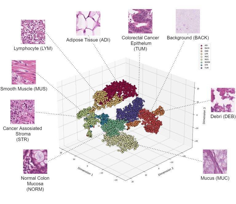
Image source: University of Jyväskylä
The new artificial neural network model has surpassed all its predecessors in classification performance. The tool has also been made freely available. The researchers published their insights in the journal Heliyon.
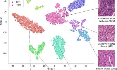
Researchers at the University of Jyväskylä, in collaboration with the University of Turku’s Institute of Biomedicine, University of Helsinki and Nova Hospital of Central Finland, have developed an advanced artificial intelligence tool for automatic analysis of colorectal cancer tissue slides. The neural network model developed in the study outperformed all previous models in the classification of tissue microscopy samples. "Based on our study, the developed model is able to identify all tissue categories relevant for cancer identification, with an accuracy of 96.74%," Fabi Prezja, the researcher responsible for the design of the method, says.
The free availability aims to accelerate future advances by encouraging scientists, developers and researchers worldwide to continue developing the tool and finding new applications for it
Fabi Prezja
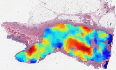
In practice, the tissue analysis involves a pathologist looking through the scanned digital microscopy slides, prepared from the patient's intestine sample, and marks, point by point, for example, where the cancerous and related tissues are visible. The tool developed in this study could save doctors' time by automating this process. The tool analyses a sample and highlights areas containing different tissue categories.The tool’s accuracy has the potential to significantly ease the workload of histopathologists, potentially resulting in faster diagnoses, prognoses and clinical insights.
The research team has made its AI tool freely available to encourage research collaboration. "The free availability aims to accelerate future advances by encouraging scientists, developers and researchers worldwide to continue developing the tool and finding new applications for it." Prezja explains.
The research team points out that despite the promising results, the introduction of AI tools to the clinical setting must be done gradually with caution. Before AI solutions can become routine clinical practice, it is essential for them to undergo a rigorous validation process to ensure that the results they produce are consistent with clinical and regulatory standards.
Source: University of Jyväskylä
03.03.2025