Image source: Adobe Stock/iQoncept
Article • Pharmaceutical research
Boosting biomarker detection with digital pathology
Digital pathology can be used to great effect in pharmaceutical research: it can accelerate analyses, give deeper insights into cellular mechanisms, and enable better understanding of their role into clinical development. This potentially offers clearer predictions on how patients may respond to treatment and lead to personalized therapies.
Report: Mark Nicholls
In a session titled “How Digital pathology can help to speed-up Biomarkers detection in Pharma Research,” Dr Carole Chotard outlined the benefits of the new technology for the field at the 10th Digital Pathology and AI Congress in London in December.
As Head of the Histopathology Department in the Servier Research Institute in Paris, France, she explained how expertise within the department covers various approaches to biomarker detection, using DNA (Fluorescent In Situ Hybridization – FISH technology), mRNA (RNAscope technology using RNA probes), and proteins (Immunohistochemistry/IHC technology using specific antibodies), which can be used to detect markers of interest on animal and human tissues. ‘These biomarkers are measurable indicators of a biological state or condition, which will have a clinical role in guiding treatment decisions. They can be predictive, prognostic, or diagnostic,’ she said.
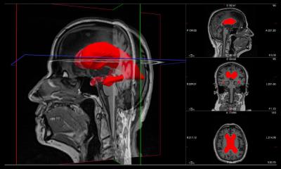
Within this, digital pathology has been key, with digital image analysis implemented in the Histopathology department to speed up quantification of biomarkers and into daily routine.
Recommended article
Article • In-depth
Focus on digital pathology
Digital pathology opens up a whole new world of possibilities in diagnosis, prognosis, and prediction of diseases. Keep up-to-date with the latest research news, medical applications, and background information on digital pathology.
Ten times the speed of a human pathologist
Dr Chotard’s presentation focused on the tumoral microenvironment in oncology and use of technology to detect several proteins of interest that could be used as biomarkers on the same tissue section (multiplex immunohistochemistry). She said there are several ways digital pathology can speed-up biomarker detection such as when performing an experiment to detect a protein on a tissue. Digitalizing the tissue slides and setting up algorithms with image analysis software can quantify the level of expression of these proteins. ‘We can then ask the computer to quantify hundreds of tissue slides. This quantification will be exactly the same for all the images and, compared to a pathologist, we will get the results much faster,’ she said.
Using Tissue Micro Arrays (TMAs) slides, which can contain several biopsies from one pathology of interest or from several pathologies, her team can perform IHC experiments and quickly screen the level of expression of a biomarker on these pathologies using automatic image analysis on digital slides. ‘That allows us to evaluate as many pathologies as we want since the computer will be ten times quicker than a pathologist to quantify the biomarker.’
A closer look at the microenvironment
A pathologist will have some difficulty to quantify by eye co-expression more than two markers in one cell whereas image analysis software can quantify dozens of markers
Carole Chotard
Immunofluorescence, used for multiplex immunohistochemistry, allows evaluation of co-expression of several markers in one cell, and calculation of distances between different cell types to understand the mechanism of action. ‘This information will help us to more easily dissect the microenvironment,’ she said. ‘A pathologist will have some difficulty to quantify by eye co-expression more than two markers in one cell whereas image analysis software can quantify dozens of markers.’
Digital pathology is being applied in various areas by Servier, including projects in oncology, immuno-oncology, immuno-inflammation and neurology but can be adapted for all types of pathologies. The aim, said Chotard, is to utilise new technologies to rapidly increase knowledge in the histopathology domain, and ‘especially to detect more and more proteins and mRNAs on a single slide. This will allow us to better understand the interaction of biomarkers within their environment.’
When working on serial sections, she said the heterogeneity of the tissues can prevent clear understanding of the interactions of proteins, especially in oncology. However, being able to observe all of them together ‘narrows down’ an understanding of the pathologies. While using the technology to characterize biomarkers and acceleration of their application into translational research and clinical development, the next step is to use AI on the histology slides to detect small abnormalities that a pathologist could not see by eye.
Paving the way for personalized treatment
For clinicians within pharma companies, improved knowledge of biomarkers expression and location will help to predict patient response to a treatment. ‘For clinicians in hospitals,’ continued Chotard, ‘the increase of digital pathology should allow small hospitals to get access to new technologies, and more knowledge in some pathologies will allow for better diagnosis.’
There are benefits for patients too. ‘In oncology, where we are the most advanced with all these new technologies and digital pathology in Servier, this will increase personalized treatment for patients, target their subtype of cancer, and evaluate if they can benefit from immunotherapy for example instead of chemotherapy.’
Profile:
Dr Carole Chotard is Head of the Histopathology Department in the Servier Research Institute in Paris, France, working from early Research programs to clinical studies. A biologist by training, she holds a PhD in Neuropharmacology from the Paris Descartes University, working in the neurosciences field using histopathology approaches. After a postdoctoral position and a role as an Investigator Scientist in Neurosciences at the National Institute for Medical Research (NIMR) in London, she joined the pharmaceutical research institute Servier in 2009 in the Histopathology section and took over the lead of the department in April 2022.
11.04.2024