Article • Going digital
Time to speed up adoption of digital pathology
Early adoption of image analytical tools and artificial intelligence are crucial if health systems across Europe are to see the full potential of digital pathology, according to a leading expert.
Report: Mark Nicholls
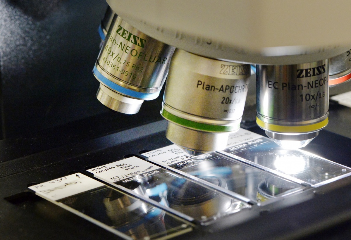
While a growing number of European institutions are beginning to embrace digital pathology, Professor Johan Lundin remains concerned about the slow pace of progress. He acknowledges that more organisations are recognising the potential of digital pathology, but several have yet not moved onto the next level of introducing analytical tools. Speaking from his experience in Finland and Sweden, he said that during the last two years major university institutions where pathology is part of the operation have acquired the microscopy scanners that are the basis for going digital and the instruments for digitalising the sample.
However, he said that the pattern in some parts of Europe was slower than he would have expected. “There are many reasons for this,” said Professor Lundin, who is Research Director at the Institute for Molecular Medicine Finland (FIMM) at the University of Helsinki. “One of the most important is that so far digital microscopy and pathology has mainly been limited to viewing. If you compare viewing a sample under a microscope, or viewing it on a screen, there is not much difference or much to gain - maybe only in speed and access to the sample archives. For the everyday work of the pathologist, I do not think they experience it as a clear improvement.”
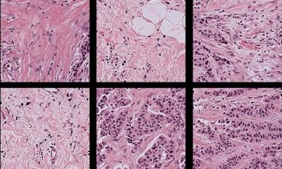
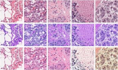
Critical to changing this, and exacerbating the implementation of digital pathology, is the addition of image analytical tools and machine learning and artificial intelligence to analyse and pre-process the samples. “With these you have a clear reason why should you go digital but without going digital you will not be able to use those tools,” he added. “Another driver is to create better interfaces where it is more convenient to view and handle the samples digitally.” He said an area where there is room for advancement is in the development of more sophisticated instrumentation with almost all instruments currently available still based on conventional technologies and typically centralised rather than located where the samples are prepared.
Pathology algorithms will reach expert levels of performance
Professor Lundin believes there are a number of steps to take to help overcome these hurdles. “When you see the huge advances in artificial intelligence applied to pathology in the last 1-2 last years, you realise there is no going back from that progress. It will definitely come, but the question is when and how fast? “We have seen from other parts of medicine - like diagnosing a melanoma from a photograph of a skin lesion or looking at the diabetic changes in the fundus of the eye – that the algorithms have reached expert levels of performance, so that means the same will of happen in pathology.
“Of course,” he continued, “it will not replace pathologists, but it will provide a number of really efficient tools to add to the clinical workflow. “What is really needed is to organise and develop the actual digitalisation process to be much more efficient and also to develop the workflow and the way pathologists interact with images and with the results from the automated analysis.”
Cost, in terms of hardware, scanners and instrumentation, remains an issue but Professor Lundin is convinced that the addition of analytical tools such as automated analysis will translate into improvements in efficiency and speed. He notes that several institutions are introducing digital pathology on a step-by-step basis; firstly by acquiring a scanner, and then digitalising archival material for teaching and seminars before leaning towards clinical samples.
A key challenge is to bridge the gap between technical researchers and clinical pathologists, one which Professor Lundin hopes the 14th European Congress of Digital Pathology in Helsinki on May 29-June 1, 2018, will help address. As joint President of the conference – which will also incorporate the Nordic Symposium on Digital Pathology – he said: “The whole topic of the conference is digital diagnostics and augmented intelligence. “I hope to gather researchers from the technical field and the clinical pathologists together to build a bridge between algorithm development and the users of these methods. “It will give the opportunity to discuss and see examples from current applications and the most recent advances in deep learning and artificial intelligence that shows the complex things you can now classify and analyse with the machine tools,” added Professor Lundin, who is also involved in the European Congress of Pathology in Amsterdam (September 2-6).
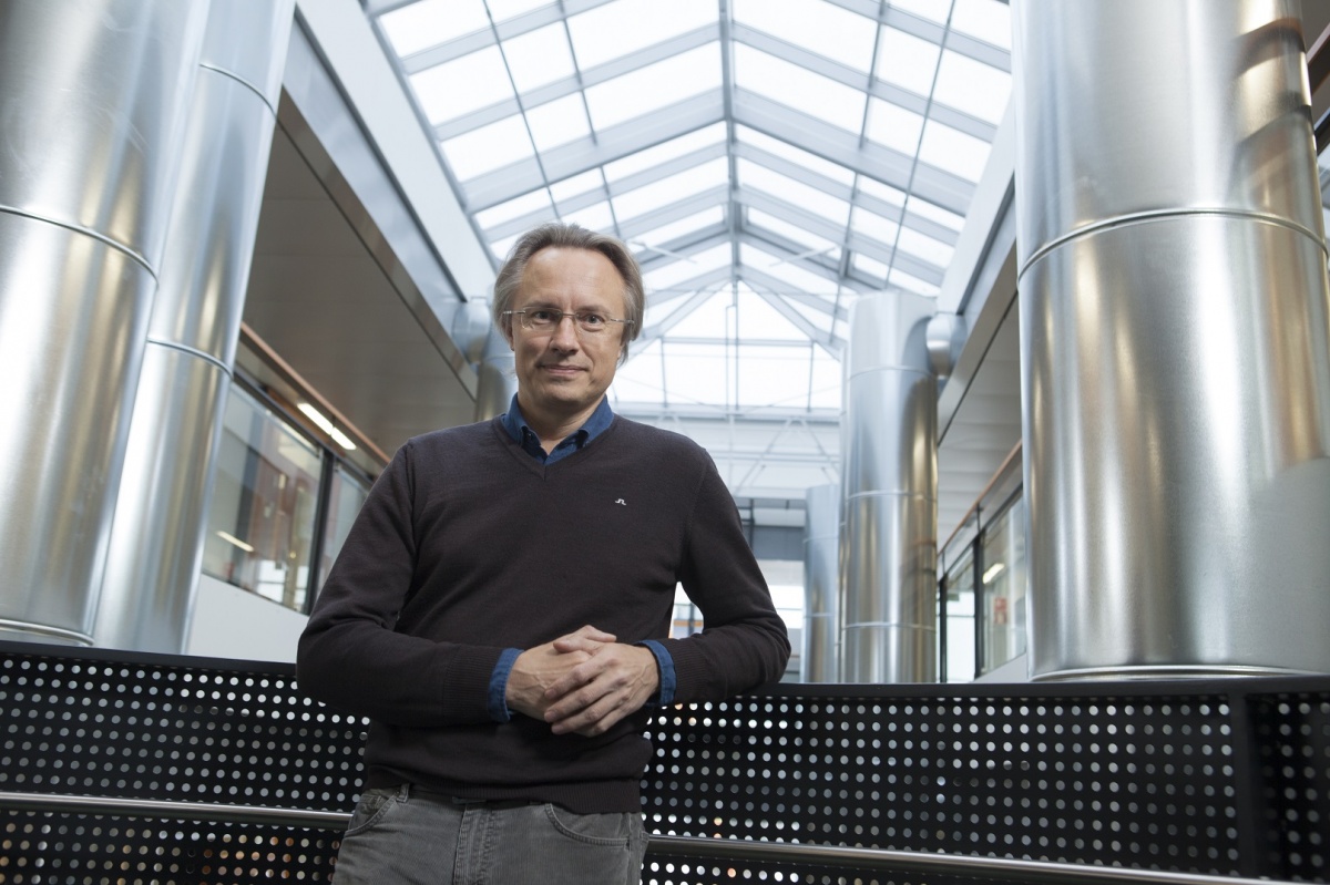
Profile:
Professor Johan Lundin is Research Director at the Institute for Molecular Medicine Finland (FIMM) hosted by the University of Helsinki and Guest Professor in Medical Technology at Karolinska Institutet, Stockholm, and also Associate Professor in Biomedical Informatics at the University of Helsinki. His key research aims are to fully utilize development within information and communication technologies for improvement of diagnostics and care of the individual patient. He is also a consultant for an academic spin-off company Finmic, which is commercialising the web microscope developed within his group.
03.09.2017