Article • Denmark
Successful digital pathology
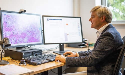
Advanced computer software underpins a service - coupled with a countrywide database, which enables Denmark’s pathologists to optimise the assessment of patients’ specimens.In turn, the digitisation of the system in recent years has led to significant improvements in pathology services, delivering greater efficiency and advances in patient safety.
Report: Mark Nicholls
Professor Ben Vainer, who will highlight the progress his country has made in this field at the Digital Pathology Congress (London. 1-2 December), believes other nations can learn from the Danish experience.
Ahead of his presentation, ‘How digitisation can improve pathology service - The Danish experience,’ he told European Hospital: ‘In large pathology labs, the high number of specimens is often a hindrance to efficient handling and ensuring patient safety without time-consuming steps, but those steps can be turned digital, releasing valuable staff resources.
‘Most important, though, is that digitisation opens up opportunities for the implementation of the new imaging techniques, which are necessary to provide each patient with the correct assessment of diagnosis and biomarker expression profile.’
Vainer, a consultant in the Department of Pathology at Rigshospitalet, at the University of Copenhagen, will focus on the Danish civil registry database and other national databases connected to this, and the use of the same laboratory information system (LIS) in all pathology departments in the entire country, with access to the national pathology database of all pathology reports in Denmark since at least 1998.
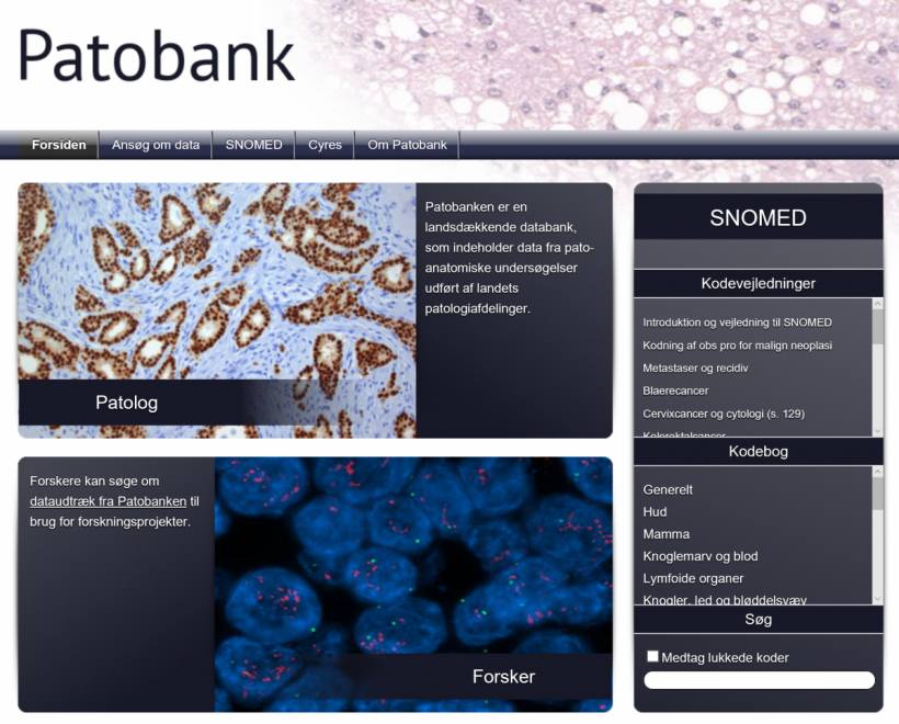
He believes this gives Danish pathologists unique opportunities to interact with each other and to study diseases from an epidemiological point-of-view.
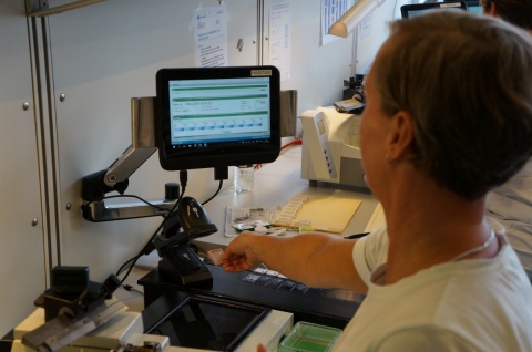
His talk will focus on the advantages of these national initiatives, giving an overview of computerisation and digitisation in routine laboratory operations, from the clinician’s ordering of a service via tissue sectioning and staining, to speech recognition and report presentation to the ordering clinician and to research-related national cancer databases.
Vainer will also discuss the important links between LIS and patient medical records in hospitals and in private practice (e.g. general practitioners), and how computerisation of the entire laboratory flow, from ordering of the pathology service to presentation of the specimen to the ordering physician, has helped ensure patient safety and eliminate time-consuming manual steps.
‘Pathology departments in Denmark have, through close collaboration, been able to build a national pathology system, where each individual pathology department serves as a sort of “branch office”,’ he added.
‘All steps of the specimens are followed through the pathology department, which gives a good global view of the departmental activities and the possibility to trace individual specimens. For the managers, this also provides good measures of operational objectives.’
The “users” – the ordering physicians – are provided with a clear overview of their patients’ specimens during the assessment process, and the patients have full access to reports on their own tissue,’ he added.
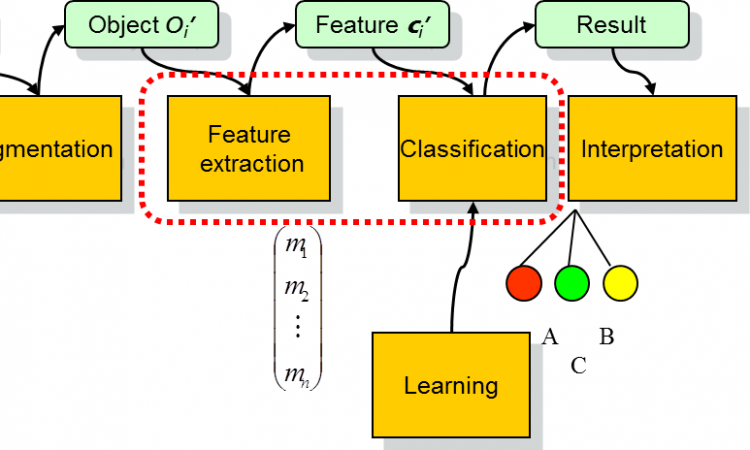
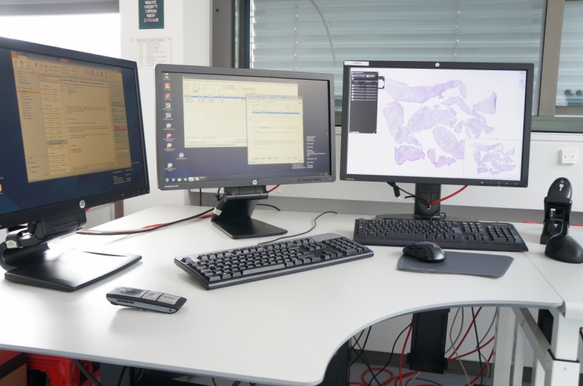
Digitisation of the laboratory processes and the link to the pathology LIS and the national pathology database opens up the opportunity for image automation, including digital image analysis and transfer of whole slide images in cases where a second opinion is needed, without compromising patient safety or the international data acts.
Patients in Denmark are also seeing clear benefits from digital pathology with a significant reduction lost specimens. ‘The risk of specimen mix-ups are minimised, and application of national pathology databases linked to national person-identification databases ensures that the pathologist always has access to previous tests performed on the patient,’ Vainer pointed out. ‘This increases the quality of the pathology assessment and hence the final diagnosis.’
The process within Denmark is constantly evolving with the introduction of the new digitisation procedures, such as automated image analysis, and substitution of conventional light microscopies with whole slide images.
‘Automated image analysis,’ he said, ‘will further increase the pathology assessment quality by eliminating subjective readings of biomarker expression, for example, in addition to eliminating the risk of patient case mix-up.’
Profile
Ben Vainer is Professor of Pathology at the University of Copenhagen and consultant pathologist at Department of Pathology, Rigshospitalet, the University of Copenhagen's main teaching hospital. His primary area of interest is digitisation of pathology in terms of education, research and diagnosing.
06.11.2016