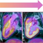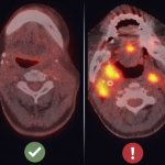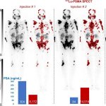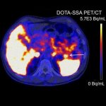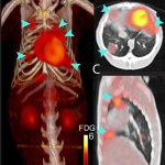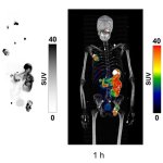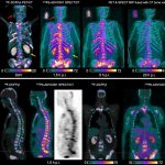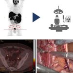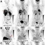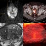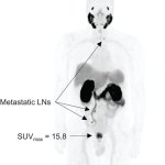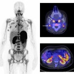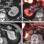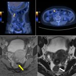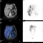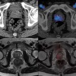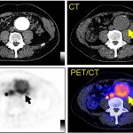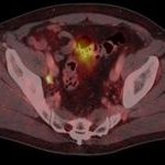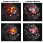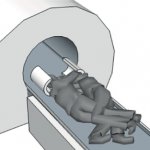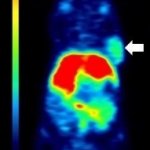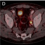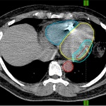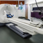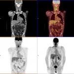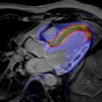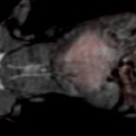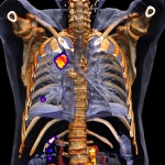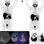
News • After an initial negative scan
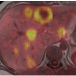
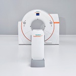
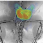
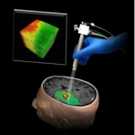
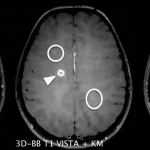
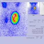
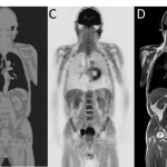
Treatment change for prostate cancer: study reveals benefit of second PSMA PET
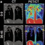
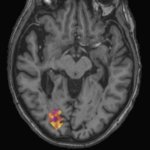
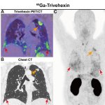
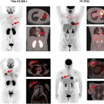
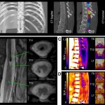
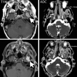
A second prostate-specific membrane antigen (PSMA) PET scan changed treatment plans for nearly half of prostate cancer patients whose first scan was negative, according to new research.