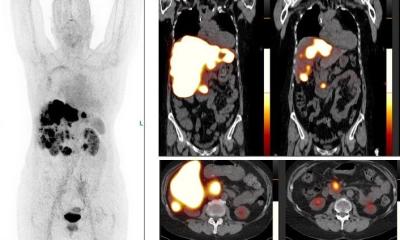
Image source: Radboudumc; adapted from: Boss M et al., JNM 2024 (CC BY 4.0)
News • Insulinoma imaging
New PET technique reveals rare pancreas tumors
A new PET scan reliably detects benign tumors in the pancreas, according to research led by Radboud university medical center.
The study in adults was published in the Journal of Nuclear Medicine. Earlier results from pediatric patients were also published in this journal.
Current scans often fail to detect these insulinomas, even though they cause symptoms due to low blood sugar levels. Once the tumor is found, surgery is possible.
The pancreas contains cells that produce insulin, known as beta cells. Insulin is a hormone that helps the body absorb sugar from the blood and store it in places like muscle cells. This regulates blood sugar levels. In rare cases, the beta cells malfunction, resulting in a benign tumor called an insulinoma. This tumor almost never spreads, but it still causes problems due to excessive insulin production, leading to low blood sugar. ‘People with this condition have little energy due to low blood sugar and often faint’, explains Marti Boss, first author of the study. ‘It's a very challenging disease. It often takes a long time before patients get a diagnosis. We can perform blood tests, but they can't confirm if a tumor is the cause or where it's located. Various scans like CT, MRI, and PET are available, but don’t always show the insulinomas.’

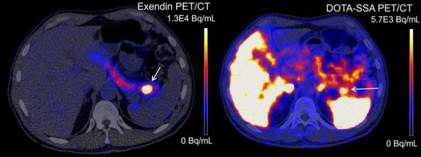
Image source: Boss M et al., JNM 2024 (CC BY 4.0)
Removing the tumor surgically resolves the problem, but first, the tumor's location must be known. Martin Gotthardt, professor of Nuclear Medicine at Radboudumc, explains: ‘In the past, surgeons would start cutting away portions of the pancreas until they found the tumor. If it was at the end, the entire pancreas would be gone. You can live without a pancreas, but you'd struggle with severe diabetes and would constantly have to manage your blood sugar. So, a better scan was urgently needed.’
Gotthardt and his team developed a completely new scan, the so-called Exendin-PET scan, which allows for the precise localization of insulinomas. They previously published results from a study in children, where the insulinoma is congenital. Now, they present findings from a study in adults, where the insulinoma developed gradually.
In the study, 69 adult patients with suspected insulinoma participated. The Exendin-PET scan detected tumors in 95% of the patients, compared to 65% with the current PET scan. When combined with CT and MRI, the current PET scan usually detected the tumor, but in 13% of cases, the insulinoma was only visible on the new scan. Boss adds: ‘We believe the new scan can replace all other scans. All the insulinomas we found with the new scan were removed, and all those patients were completely cured after surgery, even though some had been sick for decades.’
The new scan is based on a substance found in the saliva of the Gila monster, a type of lizard native to desert areas in the United States. Gotthardt explains: ‘We knew this substance specifically binds to a molecule on these tumors, the GLP1 receptor. The substance from the saliva wasn’t very stable in the human body, so we created a more chemically stable version, called Exendin. We then attached a radioactive substance to it, so it could be visible on a PET scan. Now, this mildly radioactive Exendin appears to perfectly detect insulinomas.’
The next step is to introduce the Exendin-PET scan into clinics as the standard scan for people suspected of having an insulinoma. Researchers will now assess how the scan improves patients' quality of life and how much money can be saved if other scans, like CT and MRI, are no longer needed. In addition, Gotthardt's team is investigating the potential use of Exendin for the treatment of insulinomas in a new research project called LightCure.
Source: Radboudumc
23.10.2024