Article • Cardiovascular care
Manipulating atoms and molecules with nanomedicine
Nanomedicine is set to play an increasingly important role in the future diagnosis and treatment of cardiovascular disease.
Report: Mark Nicholls
Understanding the importance of nanomedicine was enhanced by four experts who spoke at the British Cardiovascular Society conference held in June. The technology – dealing with dimensions and tolerances of less than 100 nanometres and especially the manipulation of individual atoms and molecules – is a critical component in increasingly more precise detailed approaches to cardiac care.
The speakers tackled areas such as nanomaterials for cardiovascular repair and regeneration, magnetic nanoparticles for atherosclerosis and the development of novel MRI tools to assess atheromatous plaque inflammation and stress analysis.
Professor Dave Newby spoke of ‘magnetic nanoparticles in clinical cardiovascular disease’ highlighting how magnetic resonance imaging agents have an application to cardiovascular disease, predominantly with macrophages.
Tracking active inflammation
‘Macrophages are important in lots of cardiovascular diseases – plaque rupture, heart attacks and aneurysms, for example – and resolution of injury and inflammation within that,’ said Newby, who is Professor of Cardiology at the University of Edinburgh in Scotland.
Nanomedicine and advanced imaging to study biology are currently particularly topical. ‘It’s not just body structure,’ he said. ‘It’s also about what the tissue in the body is actually doing. Nanoparticles can tell us about where there is active inflammation and where macrophages are active. ‘That can be useful because it helps us understand disease biology – where injury is happening, how diseases are occurring and how the body heals.’
Experts are using MRI, PET and other technologies to exploit the role of nanomedicine in this field as they assess arterial blockages and the disease dimension. ‘What we need to know is whether the biology is dormant, is it just going to lie there and stay unchanged for the next 10 years and never cause a problem, or is there a heart attack around the corner and what can we do to stop it happening?’
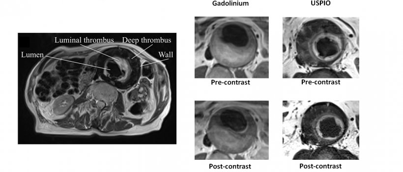
Newby outlined his work to identify ongoing inflammation using ultra-small superparamagnetic iron oxides (USPIOs) to identify hot areas within the aneurysm that are growing.
His study showed that, if the aneurysm lights up with the MR agent, it will grow bigger and surgery is necessary, or the likelihood of the aneurysm bursting increased.
Understanding cardiac injury
Newby also described how the heart heals after myocardial infarction and how, via iron nanoparticles, imaging can show how much inflammation there is in the heart and how this activity relates to the resolution and scarring of the heart attack. ‘We do not know yet whether modifying cellular inflammation will make things better or worse, because it could go either way,’ Newby pointed out. ‘If a heart does not heal well, it can burst and rupture but if it overdoes it and heals too much then you get remodelling and heart failure.’ Work with nanomedicine in this area, he said ‘are the first steps towards trying to understand how the heart is responding to injury from a heart attack.’
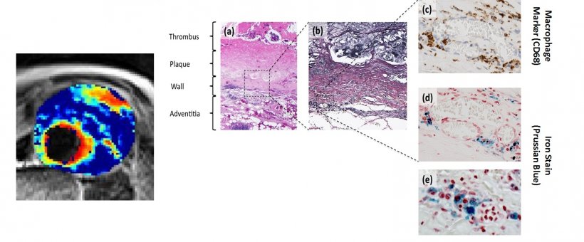
Richards et al. Circ Cardiovasc Imaging 2011;4:274-281
The speakers also included Dr Iwona Cicha, from the University Hospital Erlangen, Germany, who focused on magnetic nanoparticles for atherosclerosis - in vitro and in vivo preclinical studies. Also, Professor Patrick Hsieh, research fellow and affiliate attending surgeon at the Institute of Biomedical Sciences, Academia Sinica, Taiwan spoke of nanomaterials for cardiovascular repair and regeneration. The development of novel MRI tools assessing atheromatous plaque inflammation and stress analysis was the focus of Professor Jonathan Gillard, Professor of Neuroradiology at the University of Cambridge, United Kingdom.
Profile:
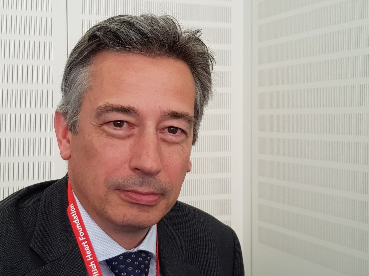
David Newby is the British Heart Foundation Professor of Cardiology at the University of Edinburgh, and Director of the Edinburgh Clinical Research Facility, plus a Consultant Interventional Cardiologist at the Edinburgh’s Royal Infirmary. His principal research interests are in advanced imaging with particular relevance to acute coronary syndromes, valvular heart disease and heart failure.
22.10.2018