Research
New Hybrid PET/MRI system improves breast cancer diagnosis
A new EU-funded project HYPMED is developing a ground-breaking imaging method for more accurate detection of breast cancer and a better, more personalised, understanding of its response to therapy.


Breast cancer is the most common type of female cancer and continues to be one of the main causes of cancer death in women. Despite the advances made in modern medicine and contemporary targeted therapies, the stage of breast cancer at the time of diagnosis is still the most important driver of patient survival. This means that there is an obvious and persisting need for an improved early diagnosis of this disease.
The project Digital Hybrid Breast PET/MRI for Enhanced Diagnosis of Breast Cancer, HYPMED for short, will develop a hybrid system of two medical imaging modalities (MRI and PET) for improved diagnosis of breast cancer and personalised therapy control. A European consortium made up of nine partners from leading universities, research organisations and industry has recently started their ambitious research initiative. “The HYPMED project combines visionary clinical expertise with excellence in physical and engineering sciences and the developed technology will greatly help us to choose an appropriate treatment that is exactly right for a given cancer in a given woman”, states, Prof. Christiane Kuhl from University Hospital Aachen and Scientific Coordinator of the project.
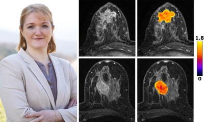
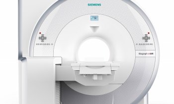
The project`s ambition is to develop a radiofrequency coil that can be connected to any regular clinical MR scanner and transform the device into a high-resolution PET/MRI hybrid system, which can be used to identify even the smallest breast cancer foci and better characterize the cancer as well as its response to therapy. Patients will also benefit as the radiation dose of the new technology will, in contrast to other PET-MRI examinations, be comparable to a regular digital mammogram.
The HYPMED approach is also likely to be transferrable to other clinical applications, such as prostate cancer detection and hybrid cardiac imaging. “We believe that this will introduce a paradigm shift in the field of PET/MR hybrid imaging with many new applications in other diseases. With the success of the HYPMED project, we will open up a whole new chapter of medical hybrid imaging,” says Prof. Schulz, head of the Department the Physics of Molecular Imaging Systems at Aachen.
The innovative concept also convinced the reviewers of the EU Horizon 2020 research program, who awarded the HYPMED project proposal the highest evaluation score possible.
Background
The HYPMED project started on January 1st, 2016 and involves, over a 4-year period, recognized organisations and industry partners from all over Europe: European Institute for Biomedical Imaging Research, AT (Project Coordinator); University Hospital Aachen, DE (Scientific Coordinator), Forschungszentrum Jülich, DE; Medical University of Vienna, AT; Technical University Delft, NL; University Hospital Münster, DE; NORAS MRI Products GmbH, DE; Futura Composites, NL; INTRASENSE, FR; Philips Electronics, NL.
The HYPMED project receives funding from the European Union’s Horizon 2020 research and innovation programme under grant agreement No 667211
Press contact
Pamela Zolda
pzolda@eibir.org
+43 533 40 64 538
www.eibir.org
28.01.2016


