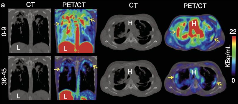
Representative coronal (left) and transverse (right) CT and 11C-PABA PET/CT images from a 33-year-old female with cystic fibrosis and confirmed M. abscessus infection in a 45-minute dynamic PET scan after an intravenous injection of 518 MBq of 11C-PABA. In the first nine minutes of the scan 11C-PABA got distributed systemically, followed by rapid clearance via heptic and renal excretion, showing accumulation of 11C-PABA only inside the affected lesion in the last 10 minutes. Not all lesions visualized on CT were PET avid, suggesting that some lesions may be sterile or had very low bacterial burden. The y axis shows time in minutes.
Image source: SNMMI; adapted from: Masias-Leon Y, Ruiz-Gonzalez C, Nino-Meza O et al., Journal of Nuclear Medicine 2025
News • Mycobacteroides abscessus
New PET technique detects hard-to-diagnose lung infections
A new PET imaging technique can accurately detect and monitor Mycobacteroides abscessus lung infections—one of the most difficult-to-diagnose conditions in patients with lung diseases.
This research, presented at the 2025 Society of Nuclear Medicine and Molecular Imaging Annual Meeting, offers a promising new diagnostic tool that could significantly improve diagnosis and treatment decisions for patients with chronic lung infections.
Mycobacteroides abscessus is a rapidly growing mycobacteria that primarily affects immunocompromised patients and those with underlying lung diseases, such as cystic fibrosis or chronic obstructive pulmonary disease. Conventional imaging tools are unable to distinguish the bacterial infection from these inflammatory diseases, and definitive diagnosis often requires invasive procedures.
“Given the constraints of conventional imaging, the development of noninvasive, bacteria-specific imaging techniques would be clinically valuable for diagnosing and selecting appropriate treatments for patients,” said Yuderleys Masias-Leon, MD, a postdoctoral research fellow at Johns Hopkins University Medical School in Baltimore, Maryland. “Our study evaluated the effectiveness of a new PET imaging approach to differentiate Mycobacteroides abscessus from the general inflammation that characterizes these lung diseases.”
This proof-of-concept data highlights the potential of 11C-PABA PET as an innovative, bacteria-specific, non-invasive and clinically-translatable diagnostic tool
Yuderleys Masias-Leon
Researchers tested the new technique—11C-PABA PET—in both preclinical and clinical settings. In vitro studies were first performed to confirm that 11C-PABA accumulated in the bacteria. Next, dynamic 11C-PABA PET was performed in a mouse model of Mycobacteroides abscessus pulmonary infection. Moving to the clinic, a 33-year-old patient with cystic fibrosis and microbiologically-confirmed Mycobacteroides abscessus pulmonary infection also underwent dynamic 11C-PABA PET.
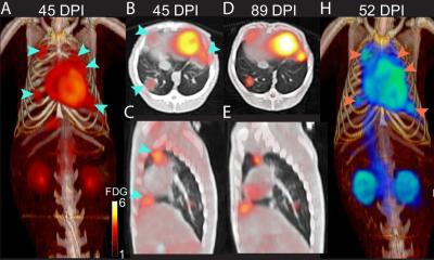
11C-PABA uptake was observed in all in vitro studies, and sustained accumulation of 11C-PABA was noted in the lung lesions of Mycobacteroides abscessus-infected mice. In the patient with cystic fibrosis, dynamic 11C-PABA PET demonstrated significantly higher PET uptake in the affected lesions compared to unaffected lung, confirming the Mycobacteroides abscessus pulmonary infection.
“This proof-of-concept data highlights the potential of 11C-PABA PET as an innovative, bacteria-specific, non-invasive and clinically-translatable diagnostic tool,” stated Masias-Leon. “In the future, 11C-PABA PET could offer rapid and accurate diagnosis to facilitate precision medicine and individualized care for patients with this infection.”
Source: Society of Nuclear Medicine and Molecular Imaging
27.06.2025