News • Physical contact research
Two people, one MRI: The science of cuddling
Researchers at Aalto University and Turku PET Centre have developed a new method for simultaneous imaging brain activity from two people, allowing them to study social interaction.
In a recent study, the researchers scanned brain activity from 10 couples. Each couple spent 45 minutes inside the MRI scanner in physical contact with each other. The objective of the study was to examine how social contact activates the brain. The results were published in the theme issue Social Interaction in Neuropsychiatry of the journal Frontiers in Psychiatry. “This is an excellent start for the study of natural interaction. People don’t just react to external stimuli, but adjust their actions moment-by-moment based on what they expect to happen next,” says Riitta Hari, emerita Professor at Aalto University.
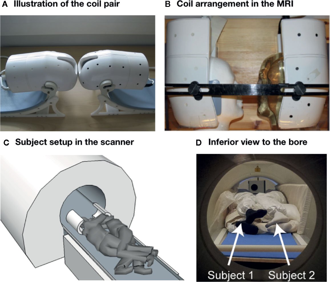
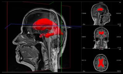
Ordinary magnetic resonance imaging is used to scan one person at a time. In the device developed at Aalto University, the head coil used for regular brain scans was divided into two separate coils. This new design allows for simultaneous scanning of two brains, when the individuals are positioned close enough to each other inside the scanner. During scanning, the participants were face-to-face, almost hugging each other. When instructed by the researchers, the subjects took turns in tapping each other's lips. Looking at the brain scans, the researchers could see that the motor and sensory areas of the couples’ brains were activated.
“During social interaction, people's brains are literally synchronised. The associated mental imitation of other people's movements is probably one of the basic mechanisms of social interaction. The new technology now developed will provide totally new opportunities for studying the brain mechanisms of social interaction,” says Professor Lauri Nummenmaa from Turku PET Centre. “For example, during a conversation or problem solving, people’s brain functions become flexibly linked with each other. However, we cannot understand the brain basis of real-time social interaction if we cannot simultaneously scan the brain functions of both persons involved in social interaction,” Riitta Hari says.
Source: Aalto University
Recommended article

News • Coital research
'Sex in an MRI scanner' – the story behind an extraordinary imaging project
This Christmas marks the 20th anniversary of the publication of “Magnetic resonance imaging of male and female genitals during coitus and female sexual arousal” in The BMJ. In its first year, it picked up the IgNobel prize for medicine, and has since become one of the most downloaded BMJ articles of all time. Dr Tony Delamothe, a former editor at The BMJ, ponders on its success.
29.04.2020