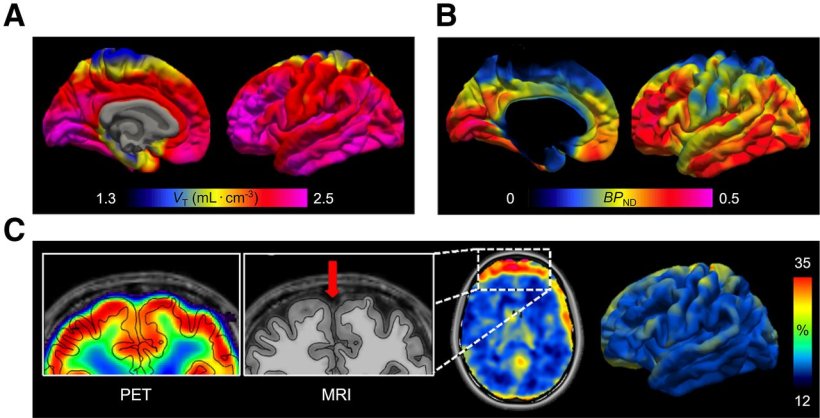
PET protein atlases for various [11C]MC1 positron PET imaging metrics targeting COX-2. (A and B) Distribution volume (VT) (A) and binding potential (BPND) (B) through volume-based analysis (left) and surface-based analysis (right) tailored to respect cortical geometry of the brain to enhance precision. (C) Between-subject variability for total COX-2 uptake, quantified as coefficient of variation, contrasting volumetric analysis (left) and surface-based analysis (right). Surface-based analysis respects the unique cortical geometry of individual participants (left), thereby reducing partial-volume effects. Moreover, a surface-based approach mitigates increased between-subject variability observed using volumetric analysis, which is exacerbated by differences in anatomy that lead to edge or boundary effects. By respecting cortical geometry, surface-based analysis reduces between-subject variability in the cortex, consequently lowering the number of participants needed in group analyses to detect similar statistical significance.
Image credit: Original image generated for this paper by Martin Noergaard, Intramural Research Program, National Institute of Mental Health, 10 Center Drive, Bethesda, MD 20892, USA; 2Department of Computer Science, University of Copenhagen, 1165 Copenhagen, Denmark. Edited by Robert B. Innis and Xuefeng Yan, Intramural Research Program, National Institute of Mental Health, 10 Center Drive, Bethesda, MD 20892, USA.
News • Nuclear imaging
PET radiotracer reveals inflammation in the brain
A novel PET imaging approach can effectively quantify a key enzyme associated with brain inflammation, according to new research
The research was published in the Journal of Nuclear Medicine. The first-in-human study, which imaged the COX-2 enzyme, offers a never-before-seen view of inflammation in the brain, opening the door for COX-2 PET imaging to be used in clinical and research settings for various brain disorders.
COX-2 is an enzyme in the brain that can be markedly upregulated by inflammatory stimuli and neuroexcitation. Researchers say that the density of COX-2 in the brain may be a biomarker and effect of inflammation, even if it is not a mediator of the inflammatory process. “While COX-2 has been widely studied in peripheral inflammation, its role in neuroinflammation has been difficult to quantify in vivo,” stated Robert B. Innis, MD, PhD, senior investigator in the Molecular Imaging Branch of the National Institute of Mental Health at the National Institutes of Health in Bethesda, Maryland. “We sought to establish a non-invasive imaging method to measure COX-2 in the living brain to enable earlier disease detection, monitor disease progression, and assess anti-inflammatory treatments.”
Neuroinflammation plays a critical role in many neurological and psychiatric diseases, including Alzheimer’s disease, Parkinson’s disease, and major depressive disorder. This could be a game-changer for personalized medicine and therapeutic development
Robert B. Innis
This study evaluated the ability of 11C-MC1 to measure COX-2 levels in the healthy human brain. First, 11C-MC1’s affinity for human COX-2 was assessed by conducting PET imaging in rats injected with lipopolysaccharide and in humanized transgenic COX-2 mice. Specific binding to human COX-2 was confirmed. Subsequently, 27 healthy human volunteers were imaged with 11C-MC1 PET to quantify the density of COX-2 in the human brain.
Among study participants, 11C-MC1 efficiently crossed the blood–brain barrier, bound to its designated target, and demonstrated high specificity for human COX-2. The radiotracer also had a moderate ratio of specific to background uptake binding potential in cortical regions. “Neuroinflammation plays a critical role in many neurological and psychiatric diseases, including Alzheimer’s disease, Parkinson’s disease, and major depressive disorder,” noted Innis. “This could be a game-changer for personalized medicine and therapeutic development. It also demonstrates the potential for developing other PET tracers to investigate neuroinflammation, broadening the applications of nuclear medicine in neurology and psychiatry.”
Source: Society of Nuclear Medicine and Molecular Imaging
02.04.2025


