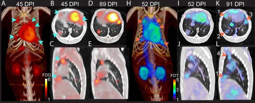
[18F]FDT scans of an Mtb-infected marmoset correlate with 4-week HRZE (combined isoniazid (H), rifampicin (R), pyrazinamide (Z) and ethambutol (E) treatment, with the number of days post infection (DPI) indicated above.
Image source: Khan RMN et al., Nature Communications 2024 (CC BY 4.0)
News • Specific TB scan
PET imaging shows tuberculosis in the body
A more accurate way to scan for tuberculosis (TB) has been developed by UK and US researchers, using positron emission tomography (PET).
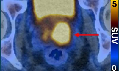
The team, from the Rosalind Franklin Institute, the Universities of Oxford and Pittsburgh and the National Institutes of Health in the USA, have developed a new radiotracer, which is taken up by live TB bacteria in the body. Radiotracers are radioactive compounds which give off radiation that can be detected by scanners and turned into a 3D image. The new radiotracer, called FDT, enables PET scans to be used for the first time to accurately pinpoint when and where the disease is still active in a patient’s lungs.
The researchers have put the new radiotracer through extensive pre-clinical trials with no adverse effects and it is now ready to go into Phase I trials in humans. Published in Nature Communications, the research was funded by the Gates Foundation and UK Research and Innovation.
With a minimum of additional training, this effective diagnostic in essence could be rolled out into most healthcare systems around the world – and most importantly, in the places where [tuberculosis] is still taking its greatest toll
Ben Davis
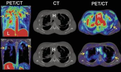
Two methods currently exist for TB diagnosis: testing for the TB bacteria in a patient’s spit or a PET scan to look for signs of inflammation in the lung, using the common radiotracer FDG. However, a spit test can show a negative long before the disease has been fully treated in the lungs, which could result in patients finishing treatment too early.
Scanning for inflammation can be helpful in seeing the extent of the disease, but it is not specific to TB, as inflammation can be caused by other conditions. Inflammation can also persist in the lung after the TB bacteria has been eliminated, leading to treatment continuing longer than necessary. The new approach developed by the researchers is more specific as it uses a carbohydrate that is only processed by the TB bacteria.
A key advantage of the new approach is that it only requires a hospital to have standard radiation control and PET scanners, which are becoming more widely available throughout the world. The new molecule is created from FDG using a relatively simple process involving enzymes developed by the research team. This means it can be produced without specialist expertise or laboratories and so would be a viable option in low- and middle-income countries with less developed healthcare systems. These countries currently see over 80% of global TB cases and deaths from the disease.
In 2021, 10.6 million people fell ill with TB and 1.6 million people died from the disease, making it the world’s second leading infectious killer after Covid-19.
FDT will enable us to assess in real time whether the TB bacteria remains viable in patients who are receiving treatment, rather than having to wait to see whether or not they relapse with active disease
Clifton Barry III
Professor Ben Davis, Science Director of the Franklin’s Next Generation Chemistry group, led the research. He said: “Finding an accurate way to identify when TB is still active in the body is not only important for initial diagnosis, but to ensure patients are receiving antibiotics long enough to kill the disease, and no longer. The common radiotracer FDG and the enzymes we’ve developed to turn it into FDT can all be sent by post. With a minimum of additional training, this effective diagnostic in essence could be rolled out into most healthcare systems around the world – and most importantly, in the places where this disease is still taking its greatest toll.”
Dr Clifton Barry III, from the National Institute of Allergy and Infectious Diseases, said: “FDT will enable us to assess in real time whether the TB bacteria remains viable in patients who are receiving treatment, rather than having to wait to see whether or not they relapse with active disease. This means FDT could add significant value to clinical trials of new drugs, transforming the way they are tested for use in the clinic.”
Source: Rosalind Franklin Institute
30.06.2024