Therapy
Molecular imaging mines deeper
The view across the Atlantic – it fills Professor Fabian Kiessling, Chair of Experimental Molecular Imaging at the RWTH Aachen (Rhine-Westphalia Institute of Technology Aachen), with optimism. The USA offers more opportunities for molecular imaging. Only recently, new tracers for Alzheimer’s were accepted as reimbursable in some centres, whilst the development of new diagnostics in Europe continues to be rather sluggish by comparison. ‘But this will change,’ Professor Fabian Kiessling is sure, ‘at the very point when we realise that many new therapies will not work without sufficient staffing, and that the use of molecular imaging will help to keep down costs and resources.’
Report: Brigitte Dinkloh

PET – gentler, faster and used intraoperatively
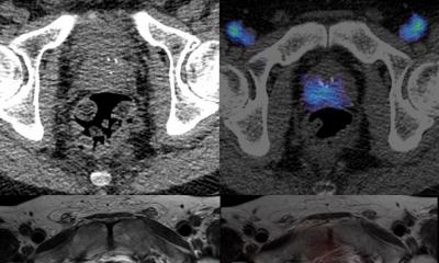
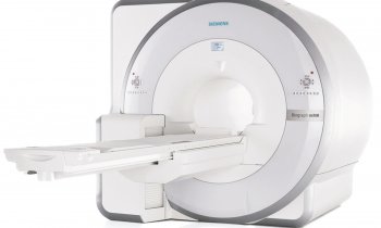
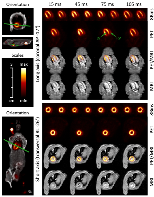
Molecular Imaging is no longer the playing field of individual researchers. To the contrary: We are now pretty certain what actually works, and there are intensive efforts to bring these applications into clinical routine. One good example of this is PET, and in particular hybrid imaging with MRI: ‘For a long time it was thought that PET has reached the zenith of its capabilities but, thanks to the new detector technology with fully digitised sensor arrays, the tracers can be detected with even more sensitivity and their spatial attribution has improved. ‘The procedure is further advanced through new geometries, such as whole-body scanners and the development of new calculation procedures. ‘This will further enhance the sensitivity of the procedure,’ Kiessling foresees. In future we will not only require significantly smaller amounts of radioactive substances but scanning will also become much faster and therefore considerably cheaper. He believes that, in the long term, MRI/PET will become fully established at least at university hospitals because of its clearly improved soft tissue contrast.
Next to PET, Kiessling also sees a big potential in Cherenkov Imaging. The idea behind this is initially to localise a tumorous lesion, or the lymph nodes, with PET imaging. During the operation a camera then enables detection of the area in vivo through light emission during the radioactive decay of the tracers. The procedure has now been developed to such an extent that the first patient examinations are being planned in New York.
Magnetic particle imaging (MPI)
The professor is particularly proud of the first ever presentation of an image from a hybrid MRI-MPI scanner. Since Bernhard Gleich first publicised the magnetic particle imaging procedure about 10 years ago there has been intensive research in this field. The principle behind it is that, in a magnetic gradient field, small iron oxides display harmonic distortions through periodic stimulation, which can be measured and which can then be utilised to carry out fast, highly sensitive imaging.
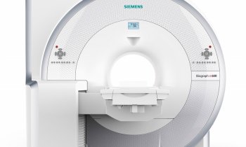
‘The Hybrid MRI-MPI scanner also allows switching between MRI and MPI. This enables visualisation of small iron oxide particles in the body with high sensitivity, which can then be hybridised into morphological MRI images. The areas of application for the hybrid scanner are still subjects of research. The first clinical MPI is located in Hamburg and will be used for cardiovascular imaging, and in particular for the fast acquisition of lung- and myocardial perfusion.’
The determination of the oxygen content in tissue, dynamically and without any additional contrast media, is a particularly interesting application of photoacoustic imaging. Kiessling: ‘It allows the detection and characterisation of tissue that is poorly supplied with oxygen, and the observation of the effects of treatment. The procedure is used in oncology, for cardiovascular disease, muscle perfusion studies and inflammatory diseases.’
The user can also inject a fluorescent dye to measure the tissue perfusion and permeability of the tissue or to detect sentinel lymph nodes. It is specifically these simple applications that should be tested a lot more because they can be quickly, clinically implemented with existing technology.
Molecular ultrasound
Kiessling is working on the discovery of new indications for the first molecular contrast medium with target-specific microbubbles. He has also achieved early measurements of perfusion and relative blood volume following the administration of only one dose of contrast medium, later obtaining molecular information on the VEGFR-2-Receptor with a further measurement. ‘In the differentiation of breast cancer we could show that molecular information is clearly superior to functional information. This is very promising and we are working very hard on the development of the clinical implementation of this procedure.’
PROFILE:
In 2008, Dr Fabian Kiessling became W3-Professor for Experimental Molecular Imaging at the RWTH Aachen. A recognised speaker from the Working Group on Methodology and Research at the German Röntgen Society (DRG) he has been a member of the European Society for Radiology (ESR) Research Board since 2011. In 2015 chaired the ESR Molecular Imaging Subcommittee. In 2014 and 2015 he also co-chaired the European Society for Molecular Imaging (ESMI) Congress and he will be Chairman of the World Molecular Imaging Congress (WMIC) in 2016.
08.07.2015


