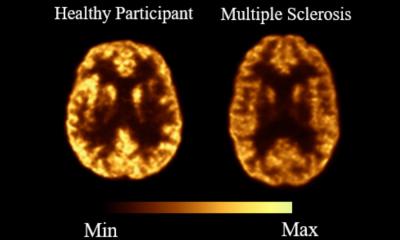
Image source: University of Turku; adapted from: Klotz L, Smolders J, Lehto J et al., Nature Medicine 2025 (CC BY 4.0)
News • Promising biomarker found
Broad rim lesions predict multiple sclerosis progression
Researchers at the University of Turku have discovered a new biomarker that can predict the progression of multiple sclerosis (MS).
The thickness of the inflammatory cell rim surrounding brain lesions was found to directly correlate with the severity and speed of disease progression. The study, led at the University of Turku in Finland by Professor Laura Airas in collaboration with German and Dutch colleagues, has been published in the journal Nature Medicine.

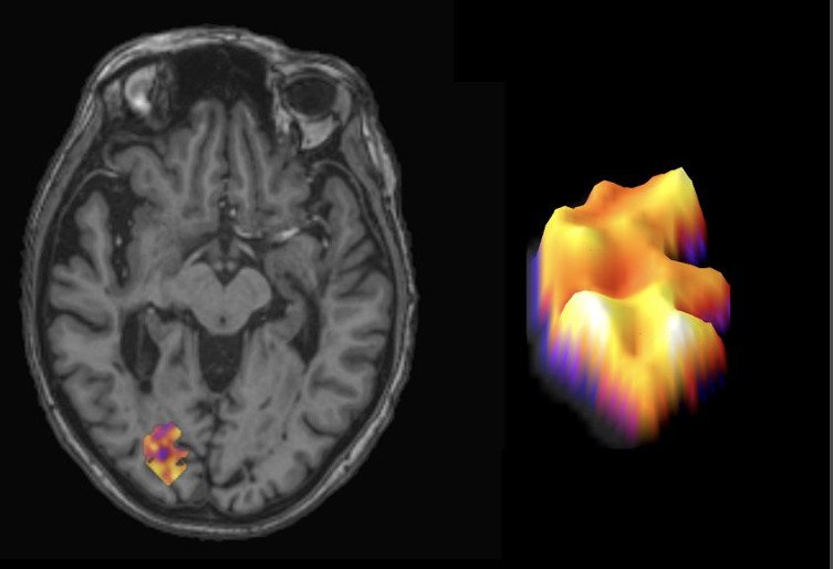
Image source: University of Turku
The research combined PET imaging data from 114 Finnish MS patients with post-mortem brain tissue analysis from Dutch MS patients. Results show that the wider the inflammatory rim around a brain lesion is, the more aggressively the disease advances. "When microglial cells form a thick rim around MS lesions, their harmful activity pushes deeper into healthy brain tissue, causing irreversible damage," says Professor Laura Airas. "This discovery allows us not only to identify patients who need more aggressive treatment earlier but also to evaluate the effectiveness of new drug candidates by observing changes in lesion rims."
The findings are expected to improve the development of treatments particularly for progressive MS, the yet undertreated form of the disease.
Professor Laura Airas is a research group leader at the InFLAMES Flagship, a joint initiative between the University of Turku and Åbo Akademi University, aiming to identify new drug targets and develop personalised therapies in collaboration with biotechnology and pharmaceutical companies. InFLAMES is part of the Flagship Programme of the Research Council of Finland.
Source: University of Turku
05.05.2025