Image source: Brugarolas et al., European Journal of Nuclear Medicine and Molecular Imaging 2022 (CC BY 4.0)
News • Neurological disease marker
Promising new radiotracer for demyelination
Loss of myelin—a protective insulating layer around neurons—is a key contributor to many neurological diseases including multiple sclerosis, traumatic brain and spinal cord injuries, stroke, and dementia; however, there are currently no imaging tests to accurately identify such demyelination and distinguish it from other pathological processes that occur in these conditions.
Investigators at Massachusetts General Hospital (MGH) have now developed such a test: a radioactive demyelination tracer that can be detected by positron emission tomography (PET) scans. They evaluated its use in humans in a study published in the European Journal of Nuclear Medicine and Molecular Imaging. “Having an imaging tool that it is specific to demyelination can help to better understand the contribution of demyelination to different diseases and better monitor a disease or the response to therapy—for example, a remyelinating therapy,” says co–lead author Pedro Brugarolas, PhD, an investigator at the Gordon Center for Imaging at MGH and an assistant professor of radiology at Harvard Medical School.
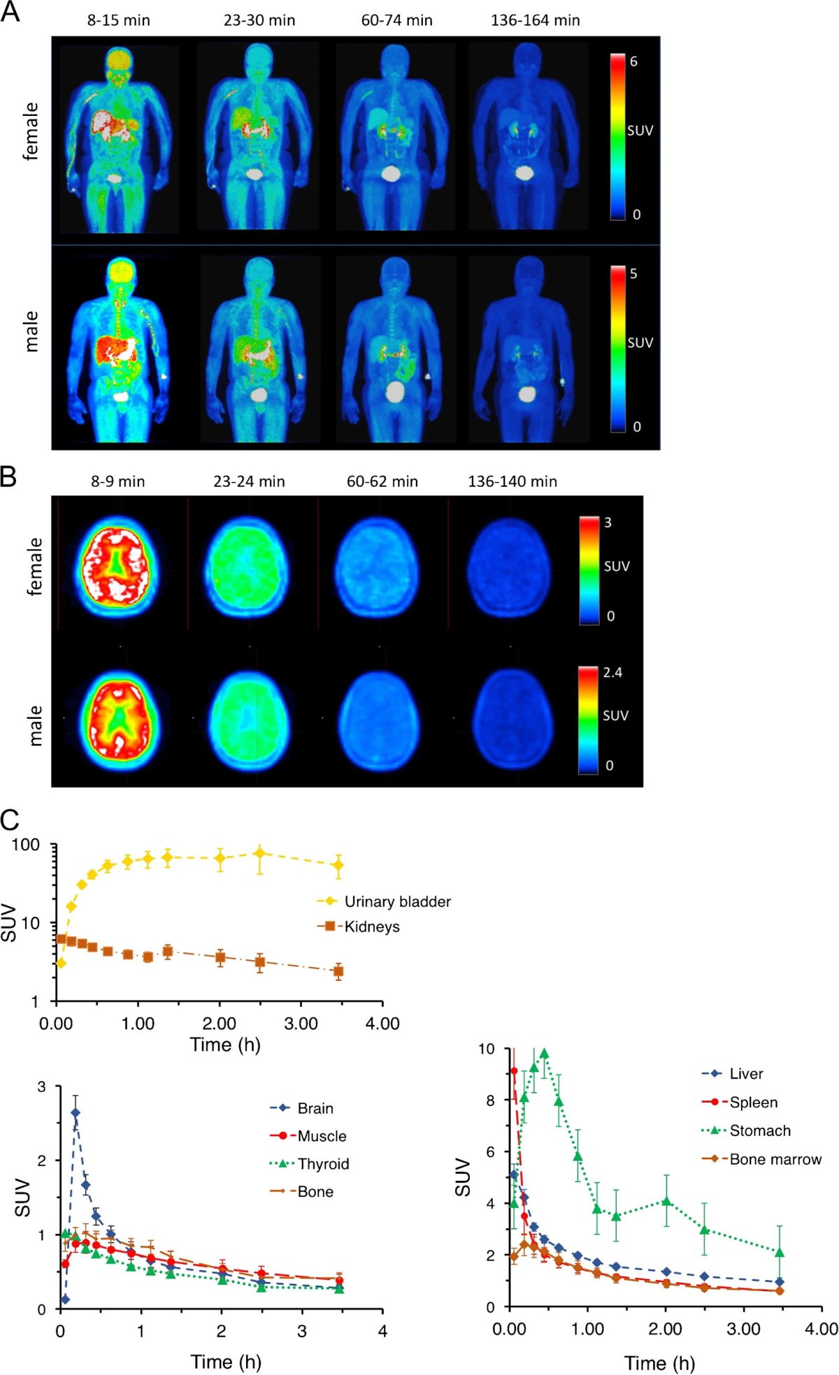
Image source: Brugarolas et al., European Journal of Nuclear Medicine and Molecular Imaging 2022 (CC BY 4.0)
In healthy volunteers, Brugarolas and his colleagues investigated the safety of their new PET tracer—called [18F]3F4AP—and looked to see what happens to it after it’s injected intravenously into the body. This was the first time the tracer was given to humans, following studies that were conducted in monkeys. The tracer distributed widely throughout the body, including into the brain, the organ of interest. In addition, the team calculated the volunteers’ radiation exposure from the tracer and they performed basic safety assessments.“
As expected for PET tracers, the radiation dose was within normal limits and we didn't see signs that could indicate that the tracer wasn't safe,” says co–lead author Moses Wilks, PhD, an assistant in physics at MGH and an instructor at Harvard Medical School. One unexpected finding was that the tracer cleared from circulation faster than expected, which they plan to investigate further.
The scientists hope that their findings will help advance research related to diseases involving demyelination. “Giving a new tracer to humans is a big deal as it requires being able to make the tracer in a sterile environment, demonstrating to the U.S. Food and Drug Administration that the trace meets a very strict quality control and getting permission from the agency to administer it to humans,” says senior author Georges El Fakhri, PhD, director of the Gordon Center for Medical Imaging at MGH and a professor of radiology at Harvard Medical School. “Once a tracer has been in humans for the first time and it has been shown to be safe—which is what this study shows—it is easier for other researchers to use it.”
Source: Massachusetts General Hospital
06.10.2022