Article • Non-invasive method
Pressure-strain loop to assess myocardial work
The value of using left ventricular pressure-strain loops as a non-invasive way to assess myocardial work was outlined in a session on cutting edge echocardiography at ESC 2022 in Barcelona. Delegates heard how the technique has benefits for cardiologists in terms of diagnosis and prognostic stratification for conditions such as heart failure, ischemic cardiomyopathy, valvular heart disease, or selecting patients for CRT (Cardiac Resynchronization Therapy).
Report: Mark Nicholls

Photo: supplied
The session provided new insights into pressure-strain loops (PSL) analysis for the non-invasive assessment of left ventricular (LV) myocardial work and its potential clinical applications. Dr Elena Galli from Rennes University Hospital in France said the objective is to present the benefits of PSL analysis as a new technique for assessment of left ventricular performance. She said: ‘The main aspect of this technique is that it allows an assessment of myocardial work in a way that is reliable and not invasive.’
Not completely precise, but very useful nonetheless
Assessment of myocardial work remains ‘a pillar of assessment of myocardial function in terms of mechanics and energy demand,’ added Galli. First introduced in the late 1970s when researchers used pressure volume loops to calculate myocardial work, it became the gold standard, but as an invasive measure it is not applicable to patients in everyday clinical practice, whereas PSL offers a non-invasive option.
The non-invasive assessment of myocardial work relies on the measure of blood pressure by an arm cuff sphygmomanometer to estimate LV pressure, and on the measure of LV strain curves by echocardiography. Pressure-strain curves are then obtained (see figure below), which allow the estimation of regional and global LV work. LV myocardial work is then assessed. ‘It is not a completely precise calculation, but it is a good estimation of myocardial work, and the regional assessment of myocardial work is shown to be directly associated with myocardial oxygen consumption assessed at FDG-PET.’
Since PSL emerged in 2012, it has been validated by various research groups for estimating myocardial work in a reliable fashion in several different pathologies, she added, and most widely shown as beneficial in CRT candidates. ‘Studies also show applicability of the method to other clinical conditions; for instance, in patients with hypertension, systolic heart failure, diastolic heart failure, and valvular heart disease, particularly aortic stenosis,’ said Galli. ‘Also, there are some pioneer studies that have started for the non-invasive assessment of the myocardial work of the right ventricle by the analysis of pressure-strain loops.’
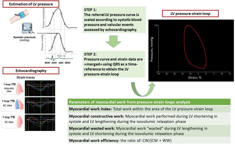
Image source: Dr Galli
Ready for clinical practice
Other approaches to assess left ventricular function and performance – such as left ventricle ejection fraction and strain – have limitations, she continued, because they simply measure LV fiber shortening and do not take into account the effect of afterload on LV function. As such, PSL seems to provide a more precise and physiological assessment of LV function. ‘This is one of the main points of the technique, together with its non-invasive nature,’ she said.
In the future [PSL] could replace some of the parameters that we use now, for instance LV strain
Elena Galli
Galli believes cardiologists can use PSL to highlight local differences in myocardial work to enable selection of patients receiving CRT and, in the future, it may offer pathophysiological insight into other disease states, such as heart failure and valvular heart disease. The technique is only slowly entering clinical practice and not being widely applied to patients despite the increasing numbers of studies being conducted in the field. ‘We can see that the evidence in research, and interest on parameters derived by PSL analysis, is increasing,’ Galli added. ‘I think that in the future it could replace some of the parameters that we use now, for instance LV strain, but we need wider evidence to prove it, so we are still at the starting point of the method.’
Profile:
Dr Elena Galli is a cardiologist and researcher at the University Hospital of Rennes, France, and INSERM where her main clinical and research interests concern cardiovascular imaging, particularly echocardiography in several different modalities. She is a Fellow of the European Society of Cardiology, a board member of the French Society of Cardiovascular Imaging, and serves on committees of the European Association of Cardiovascular Imaging.
26.08.2022