Article • PET/MRI, PET/RF & more
Disruptive innovations in molecular imaging
Molecular imaging is an exciting field for scientists who are willing to explore and innovate, prominent Spanish physicist José María Benlloch pointed out when he reviewed some of the most impacting and recent innovations in his portfolio during a meeting in Valencia.
Report: Mélisande Rouger
‘Our mission is to develop innovative sensitive and harmless medical imaging instruments for early detection of diseases and follow-up. We also work to create new minimally-invasive therapies based on physics mechanisms, and enable technology transfer to the industry,’ said Benlloch, Director of the Institute for Instrumentation in Molecular Imaging (I3M). Of late, Benlloch and team developed the first PET scanner for preclinical evaluation of small animals – a technology they transferred to Bruker, and which Stanford University now uses.

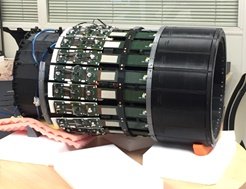
Another recent ground-breaking innovation is the PET/MRI scanner dedicated to brain examination developed by I3M within the MINDVIEW (Multimodal Imaging of Neurological Disorders) European Project. The project aims to diagnose and treat schizophrenia, severe depression, and all mental disorders. The new, high resolution PET/RF coil for simultaneous PET/MRI acquisition was originally designed for 7-T MRI, but soon upgraded to 9-T and 15-T at different sites across the world. Benlloch believes this has tremendous commercial potential. ‘There are about 40,000 MR scanners in the world, so the idea was simply to replace the radiofrequency device designed for brains to have an MR examination that also offers PET. Most MR examinations – perhaps over 40% – are for brain examination, so it makes sense to have such a solution,’ he pointed out.
While working on the new machine, researchers found it must be adapted to accommodate a very large and heterogeneous population of patients. For example, the elderly with Alzheimer’s may be challenging to image, due to age and condition. In addition, many improvements must be made, starting with compatibility of the newly developed sensors with MR. ‘These are silicon sensors that do not contain nickel, not to interfere with the magnetic field. Many of these sensors failed but images were obtained despite those errors. However, using appropriate software, the faults became almost undetectable. We even managed to obtain resolution lower than 2mm, which posterior solutions were unable to achieve,’ Benlloch explained.
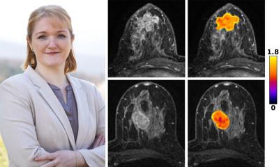
A while ago, I3M also created the innovative PET mammograph and new molecular compounds for early breast cancer diagnosis and evaluation of chemotherapy response, as part of the EU Mammography with Molecular Imaging (MAMMI) project. The Mayo Clinic in Rochester, USA, installed the MAMMI Breast PET scanner for primary systemic neoadjuvant therapy, with excellent results. ‘We could clearly see tumour extension reduction and reduction of glucose uptake after the first chemotherapy cycle,’ Benlloch said. About 30 models of the PET mammograph were then sold.
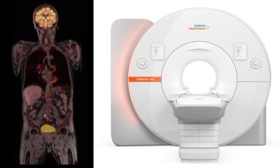
The EXPLORER total-body PET scanner, released in 2018 and originally conceived by Simon Cherry and Ramsey Badawi from UC Davis, is an inspiring advance. ‘The Explorer has produced exceptional images. I have never seen such high-quality images in a commercial setting,’ Benlloch confirmed. Chinese radiologists have widely purchased the scanner, which enables image-capture of the human body in under a second. However, Western counterparts have not shown the same enthusiasm yet; they tend to prefer big scanners that can accommodate every patient, even though these are not cost-effective enough for private practice, Benlloch believes. ‘We are going to develop something to interest everyone, i.e. a scanner that can accommodate every patient and has a high turnover – imaging many patients in an hour.’
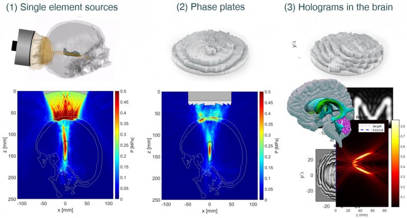
Having a solution that optimises transcranial ultrasound propagation for blood-brain barrier opening can be an interesting development for brain applications. Benlloch and team recently patented such a device, which works by applying high-intensity focused ultrasound (HIFU) to a membrane created with a 3-D printer using CT or MR images. The difference with HIFU is that the solution only uses one ultrasound transducer. ‘We mold the crane based on information from a CT or MR scan. We create the membrane, with a 3-D printer, to highlight which brain area we want to focus on with ultrasound. This could be helpful for neurostimulation,’ he pointed out.

MRI is excellent to image soft tissues but not so much for hard tissue, such as teeth or bone. The MRI lab at I3M therefore developed MR technology for simultaneous visualisation of both soft and hard tissue in dental applications. ‘Teeth produce a signal on MR, provided one uses a sensor that is fast and sensitive enough to capture that signal. Our solution may be even better. We change the magic angle of MR by rapidly oscillating between tissues.’ The next frontier will be to image the bone, jaws and crane indifferently, and MR has already proven it could get there using special sequences.
The I3M has also given birth to innovations in MR image-guided therapy, to drive magnetic nanoparticles to biological tissues using external magnetic fields and guided by MR; and vortex elasticity imaging, using acoustic vortices. The institute has also pioneered MRI development at the cellular range, with non-invasive visualisation of individual human cells in vivo (< 10 μm) and in real-time, and in vivo mouse brain imaging with 29 μm resolution at 15.2-Tesla. Last but not least, the group is also active in software and biomarkers development.

Profile:
José María Benlloch Baviera is Professor of Physics at the National Spanish Research Council (CSIC) and Director of the Institute for Instrumentation in Molecular Imaging (I3M) in Valencia, Spain. He worked at the Massachusetts Institute of Technology (MIT), under the aegis of 1990 Physics Nobel Prize winner Jérôme Friedman. Benlloch has published more than 290 articles in scientific reviews, coordinated around 30 investigation projects and holds 15 patents. He has also created three spinoff biomedical engineering companies.
26.02.2019