Image source: IOCB Prague
News • Macrocyclic hybrid probe
New dual PET/MRI contrast agent to enhance kidney diagnostics
A research team from IOCB Prague, working in collaboration with the University of Tübingen, Germany, and the Faculty of Science, Charles University, has developed a new type of contrast agent that can be used in both magnetic resonance imaging (MRI) and positron emission tomography (PET).
This breakthrough resolves a major issue that previously hindered the integration of these two imaging techniques into a single device. Their discovery has the potential to significantly enhance diagnosis and subsequent treatment, particularly for kidney diseases and tumors. The research was published in Angewandte Chemie.
We are on a path that will eventually allow us to determine not only what disease a patient has but also the stage, type, and aggressiveness of the condition
André Ferreira Martins
For a long time, medicine has been searching for new ways to refine and streamline available imaging methods. The ultimate goal was to combine the benefits of MRI and PET. While MRI is exceptional for imaging internal organs and tissues with high water content, PET can image even very small amounts of matter, making it capable of sensitively detecting molecular markers, for instance, in cancer cells. The fusion of both techniques was challenging because the strong magnetic field in MRI complicates the functioning of the electronics required for PET. However, this issue has been overcome, and hybrid PET/MRI machines are appearing in hospitals as the most advanced imaging technology. The remaining challenge was the development of a dual-purpose contrast agent that would work with PET and MRI simultaneously. The collaborative discovery by Czech and German researchers may be the solution.
Image source: IOCB Prague; photo: Tomáš Belloň
The described approach is simple and effective. Dr. Miloslav Polášek, head of the Coordination Chemistry group at IOCB Prague, where the work started, explains: “Previous attempts to make PET/MRI probes aimed to achieve advanced and specialized functions, which led to complicated molecules with difficult synthesis and a narrow scope of use. We turned this around by designing a molecule that is simple to use and has broad applicability, one that radiologists will intuitively know how to use. It replicates all the favorable properties of current clinical MRI contrast agents and also provides a PET signal. This adds another dimension of information, improving accuracy and opening new diagnostic applications.”
Recommended article
Article • Cancer imaging
PET/MRI offers significant patient benefits
PET/MRI is offering new imaging opportunities for cancer patients at various points along the care pathway with its ability to assess different biological processes and its increased specificity.
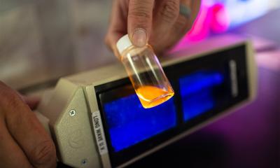
“Our solution is a cleverly designed molecule that combines gadolinium and radioactive fluorine-18, which is commonly used in medical scans and is easy to obtain. Connecting these two parts required overcoming several problems, including the large difference in the amounts needed for MRI and PET images. We solved this by using an innovative technique to swap nonradioactive fluorine atoms in the MRI contrast agent for radioactive fluorine-18 atoms. The reaction is elegant, fast, and effective. In less than 30 minutes, we can use automated synthesis to prepare enough of this agent to treat five patients,” says Dr. Jan Kretschmer, one of Dr. Polášek’s former PhD students and the first co-author of the study, who currently works at the Werner Siemens Imaging Center at the University of Tübingen in the group of Prof. André Ferreira Martins.
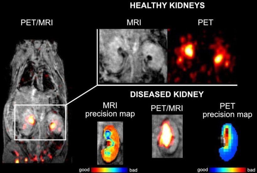
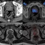
Prof. Martins describes a potentially groundbreaking discovery made during testing in a mouse model: “Unexpectedly, we found that one seemingly healthy mouse had kidney problems. The right kidney showed systemic filtration patterns that could only be detected with the simultaneous detection of PET and MRI. This approach enabled us to monitor noninvasively, in real time, the biochemical behavior, distribution, and accumulation of the probe in living, healthy subjects, thereby explaining its unexpected behavior. This method represents a pioneering step toward personalized diagnostics, showcasing the significant diagnostic potential of our hybrid molecule. This is a revolutionary discovery in the field of precise imaging. We are on a path that will eventually allow us to determine not only what disease a patient has but also the stage, type, and aggressiveness of the condition.”
The new hybrid contrast agent is patented, and researchers are seeking potential investors. According to Dr. Polášek, this compound, originating from IOCB Prague, has properties that make it the first serious candidate for a PET/MRI agent usable in a clinical setting.
Source: Institute of Organic Chemistry and Biochemistry of the Czech Academy of Sciences
06.08.2024