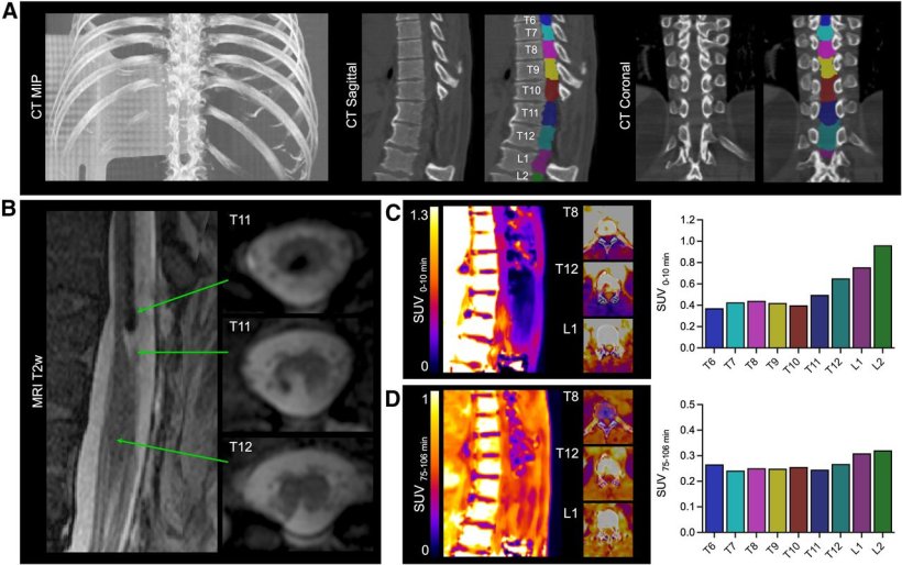
[18F]3F4AP in a human subject after mild incomplete spinal cord injury. (A) Maximum-intensity projection (MIP), sagittal, and coronal CT images showing laminectomies at T10–T12. Selected regions of interest for segmentation of spinal cord are shown for both sagittal and coronal views. (B) T2-weighted (T2w) MR sagittal and axial images showing spinal lesion. (C) Early PET sagittal images showing high PET signal in cord around injury and quantification 0–10-min SUV at selected spinal segments. (D) Late PET sagittal images showing increased PET signal in cord around injury and quantification of 75–106-min SUV at selected spinal segments. For both early and late PET, axial images at T8, T12, and L1 are shown.
Image source: Ramos-Torres KM, Conti S, Zhou YP et al., Journal of Nuclear Medicine 2025 (CC BY 4.0)
News • Potential for recovery
PET tracer offers new insights into spinal cord injuries
A novel PET technique that visualizes spinal cord injuries provides critical information about which patients may be able to regain mobility, according to new research.
This is according to new research published in the February issue of The Journal of Nuclear Medicine. By detecting intact nerve connections in the injured spinal cord, a newly developed radiotracer has the potential to help diagnose injuries more precisely, monitor recovery, and evaluate the effectiveness of new therapies in clinical trials.
Each year, approximately 18,000 Americans suffer from spinal cord injuries. These injuries can result in devastating loss of mobility, often causing lifelong struggles to regain independence and quality of life. During the initial months after injury, there are rapid degenerative changes, such as demyelination at and around the lesion, which are associated with a less favorable recovery outcome. “Recovery outcomes for spinal cord injuries can be highly unpredictable, and current imaging techniques don’t always provide clear answers about who might recover,” said Clas Linnman, PhD, assistant professor at Spaulding Rehabilitation Hospital and Harvard Medical School in Boston, Massachusetts. “Our study investigated whether PET imaging could detect spared nerve connections in the injured spinal cord that may indicate a better chance of recovery.”
This study underscores the incredible potential of molecular imaging to investigate complex neurological challenges, such as spinal cord injuries, multiple sclerosis, traumatic brain injury, and even some dementias
Pedro Brugarolas
A novel PET tracer, 18F-3F4AP, was developed to image demyelinated axons (damaged nerve fibers) in spinal cord injuries. In a preclinical research model, rats with incomplete spinal contusion injuries were imaged with 18F-3F4AP PET at multiple time points up to one month after injury. PET imaging results were then compared with autoradiography and immunohistochemistry of postmortem spinal cord tissue. Additionally, a proof-of-concept study of 18F-3F4AP PET in two human patients with spinal cord injuries of different severities was performed.
18F-3F4AP PET of rats revealed a more than two-fold increase in tracer binding at the injury site at seven days after injury relative to baseline. Autoradiography, histology, and immunohistochemistry confirmed 18F-3F4AP’s targeting of demyelinated axons. In humans, 18F-3F4AP differentiated between a severe and a largely recovered incomplete spinal cord injury, indicating axonal loss and demyelination, respectively. Moreover, alterations in tracer delivery were evident on dynamic PET images, suggestive of differences in spinal cord blood flow between the injuries.
“This study underscores the incredible potential of molecular imaging to investigate complex neurological challenges, such as spinal cord injuries, multiple sclerosis, traumatic brain injury, and even some dementias,” noted Pedro Brugarolas, PhD, assistant professor at Massachusetts General Hospital in Boston. “Such a tool could revolutionize how we diagnose, monitor, and understand neurological diseases, paving the way for advancements in molecular imaging and nuclear medicine.”
Source: Society of Nuclear Medicine and Molecular Imaging
25.02.2025