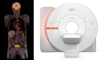
Article • FDG-PET imaging of the brain
The nuclear medicine approach to Alzheimer’s
Nuclear Medicine techniques have an important role in the clinical diagnosis of patients with cognitive impairment. And such techniques are not only valuable in a clinical setting but also in research, according one of the leading experts in the field, Javier Arbizu, who is Professor of Radiology and Nuclear Medicine at the Faculty of Medicine at the University of Navarra, Spain.
Report: Mark Nicholls
Image source: Shutterstock/Naeblys

He will expand on this in his virtual presentation to ECR, “PET as part of the biomarker toolbox for early clinical diagnosis of Alzheimer's disease” on July 16. Key objectives of the broader session, under the title of “Alzheimer's disease and neurodegeneration: visualising the invisible”, are to appreciate that dementia is more than Alzheimer's, understand that neurodegenerative diseases are overlapping and may co-exist, and to acknowledge the need for early and specific detection of cognitive decline for future clinical trials.
Speaking to Healthcare in Europe ahead of ECR, Professor Arbizu specifically outlined how he will discuss the different radiotracers available to evaluate the specific processes involved in the neurodegenerative conditions found in dementia; throw light on the benefit of PET molecular imaging in the diagnostic workup of Alzheimer's disease (AD); and help delegates to appreciate the role of different PET and SPECT techniques in the differential diagnosis of neurodegenerative disorders.
Professor Arbizu, who is also Consultant at the Nuclear Medicine Department of the Clinica Universidad de Navarra in Pamplona, said: “The two pathological hallmarks of Alzheimer's disease can currently be seen in vivo using different radiotracers. On the one hand, amyloid plaques start accumulating in the brain of the patient years before the onset of clinical symptoms, and later on tau neurofibrillary tangles spread in the brain just before the cognitive complains. In addition, we can obtain tomographic images of the neuronal dysfunction or neurodegeneration using 18F-Fluorodeoxyglucose (FDG-PET), as well as presynaptic dopaminergic system, which are affected in other conditions. Interestingly, all these processes involved in dementing neurodegenerative conditions appear before structural abnormalities like atrophy can be observed.”
The main benefit of PET molecular imaging in the diagnostic workup of Alzheimer's disease, he continued, is to help clinicians (neurologists, psychiatrists or geriatricians) to define the aetiological diagnosis at the early stages of neurodegenerative diseases, particularly when clinical diagnosis using the standard tools is uncertain. He also pointed to the National Institute of Aging-Alzheimer’s Association NIA-AA 2018 research framework for AD, which proposed a biological definition of AD based on the presence of amyloid and tau biomarkers, with cognitive symptoms and neurodegeneration biomarkers (FDG-PET) used to stage severity.
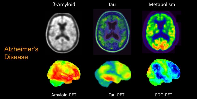
The role of different PET and SPECT techniques in the differential diagnosis of neurodegenerative disorders can be defined and influenced by the patient’s clinical profile and the specific uncertainty of the clinician. Professor Arbizu explained: “When AD is the suspected clinical diagnosis, amyloid imaging will be the first option to rule-out AD and provide specific information of the underlying pathology. However, when other than AD conditions are also suspected probably FDG-PET or even tau-PET will be more useful at the beginning of the diagnostic workup. When the cognitive decline is associated with a parkinsonian syndrome, molecular imaging of presynaptic dopaminergic system can help to rule out AD.”
In my opinion, PET molecular imaging is particularly useful in the prodromal stages of the diseases, when only mild symptoms are present
Javier Arbizu
Professor Arbizu believes PET imaging using different biomarkers is increasing in relevance as an important complementary tool in the diagnostic workup of patients with cognitive impairment. “In my opinion,” he added, “PET molecular imaging is particularly useful in the prodromal stages of the diseases, when only mild symptoms are present.” He also noted that while PET molecular imaging is a clinically-available technique using commercialized radiotracers, the development of new PET biomarkers is ongoing, particularly new generation of tau-PET tracers and those related to neuroinflammation and synaptic activity could prove relevant in the understanding of mechanisms involved in dementia.
Key take home message for delegates are that it is feasible to image in vivo specific neuropathological aspects of dementing neurodegenerative conditions by means of nuclear medicine tools; PET biomarkers have improved the understanding of disease pathophysiology of molecular pathologies, as well as their interaction, and their functional consequences; and in the clinical setting, amyloid, tau and FDG-PET combined with structural imaging helps the clinical diagnosis of the main cause of dementia (AD) and its differential diagnosis at the very early stages.
Profile:
Javier Arbizu is Professor of Radiology and Nuclear Medicine at the Faculty of Medicine at the University of Navarra, and Consultant at the Nuclear Medicine Department of the Clinica Universidad de Navarra in Pamplona, Spain. He is currently involved in different international clinical trials and research projects. He is coordinator of the PET ADDS Consortium (Spanish registry of amyloid PET studies), studying the clinical value of amyloid-PET in the diagnosis of subjects with cognitive impairment and the influence in the treatment prescription; the role of FDG-PET in the prediction of progression to AD dementia in MCI subjects; and FDG-PET patterns in different neurodegenerative parkinsonian syndromes.
15.07.2020