

Article • Cinematic Rendering in surgery
3D visualisation is like ‘body cinema’
A physician has basically two possibilities to look inside a body: putting a scalpel to the patient’s skin or using an imaging device. Both options have advantages and disadvantages.
Report: Wolfgang Behrends
Surgery Department of University Hospital Erlangen
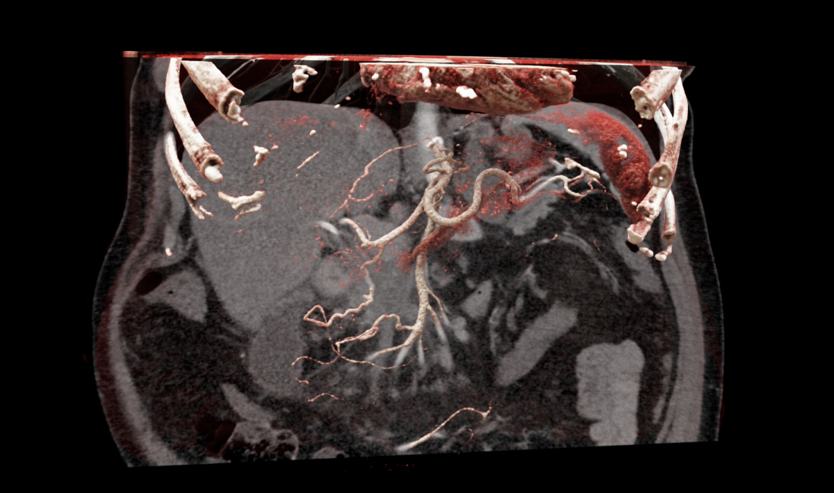
Now, Siemens Healthineers has combined the advantages of both methods and developed Cinematic Rendering, a new 3D visualisation approach. The name already points to the roots of the technique – the cinema. But it does much more than providing impressive images.
The human body is a marvel, the anatomy highly complex. “This complexity is difficult to grasp, particularly in the OR,” says Dr Christian Krautz. The surgeon is heading the current clinical evaluation of Cinematic Rendering for surgery purposes at University Hospital Erlangen, Germany. The photo-like 3D display of conventional DICOM images that the new procedure offers, allows quick orientation inside the body. “The technique makes it much easier for us to get an overview of individual anatomy,” says Dr Krautz. “That’s like body cinema. Even seasoned surgeons are wowed – and it is a real help for our brains,” seconds Professor Dr Robert Grützmann, Director of the Surgery Department.
Better overview in difficult situations
But how do spectacular images contribute to positive surgery outcomes? “Take liver surgery, for instance,” says Christoph Löhr, Senior Manager Strategy at Siemens Healthineers, “The liver is a highly perfused organ surrounded by a thick layer of fatty tissue. Interlinked and adjacent vessels are visible neither with the naked eye nor on images. Even experienced surgeons have to use the combination of years of experience and haptics: they feel the vessels with their hands.” But things are easily overlooked – or rather ‘overfelt’ – with potentially fatal consequences for the patient. Thus, 3D visualisation of the layout is a huge help. “This is exactly what Cinematic Rendering does per mouse click,” says Löhr. One of the main design ideas was to keep the operating concept of the software minimalistic: filter functions can show and hide bones, vessels and tissue. This provides images that immediately show clinical issues.
“Democratisation of the tumour board”
In the clinical study, cases are examined retrospectively in order to test the new procedure. Löhr: “Surgeons see 2D DICOM images as well as Cinematic Rendering images and are asked how they would decide based on these two types of images. We are particularly interested in complex cases such as pancreas or liver. These are organs where we expect the new technology to offer the greatest value-add and more security.”
The developers are confident that there are many more areas where Cinematic Rendering will prove its mettle. “We see enormous potential in teaching, in patient communication and in forensics,” explains Dr Klaus Engel, Principal Key Expert at Siemens Healthineers and inventor of Cinematic Rendering. “Moreover, tumour boards will benefit from the new technology because the images are easier to interpret. This will lead to a democratisation of the boards, faster agreement and hopefully to better decisions for the patients,” Dr Grützmann adds.
Sophisticated algorithms for real-life applications
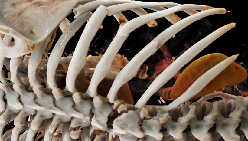
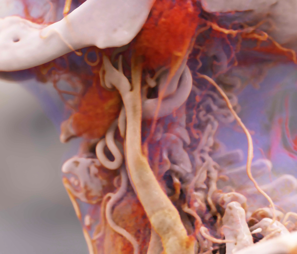
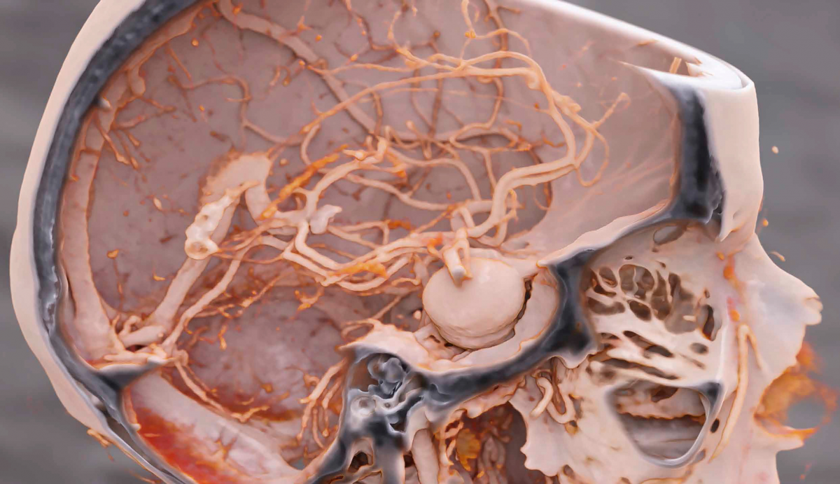
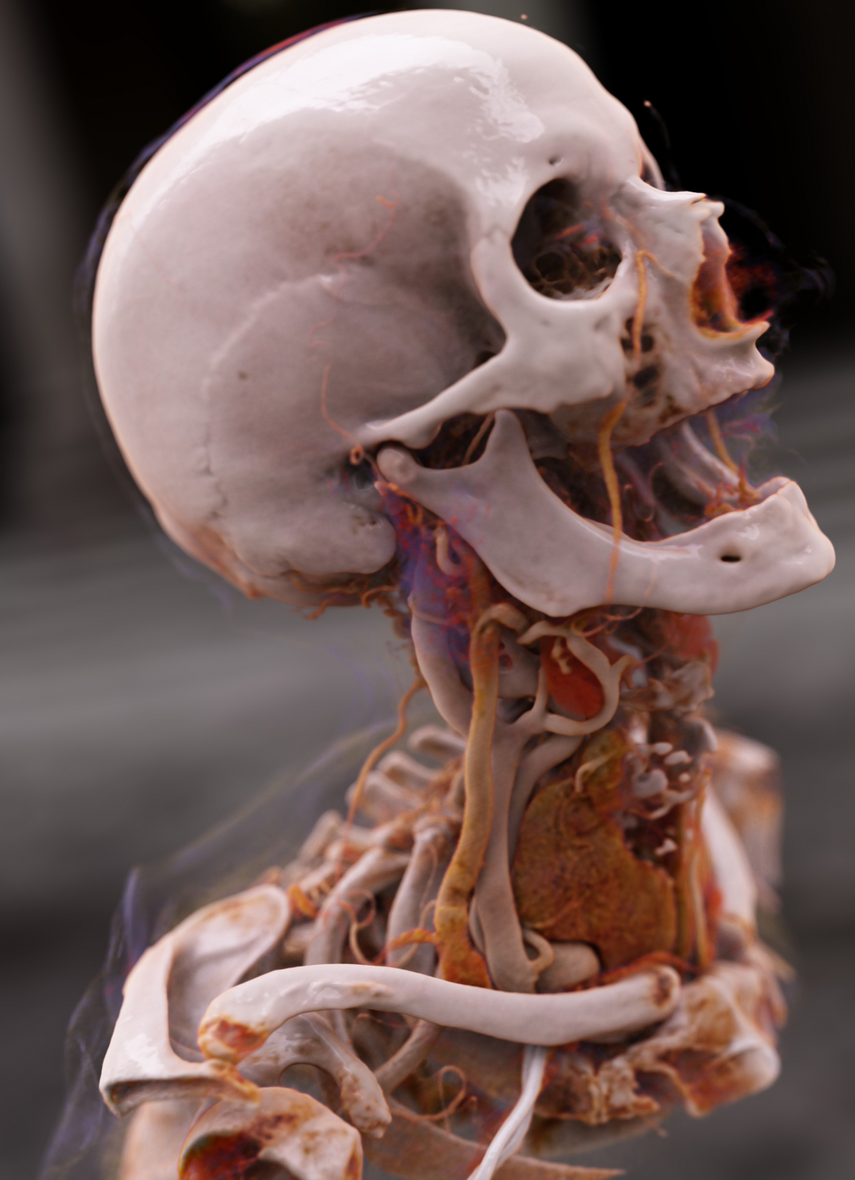
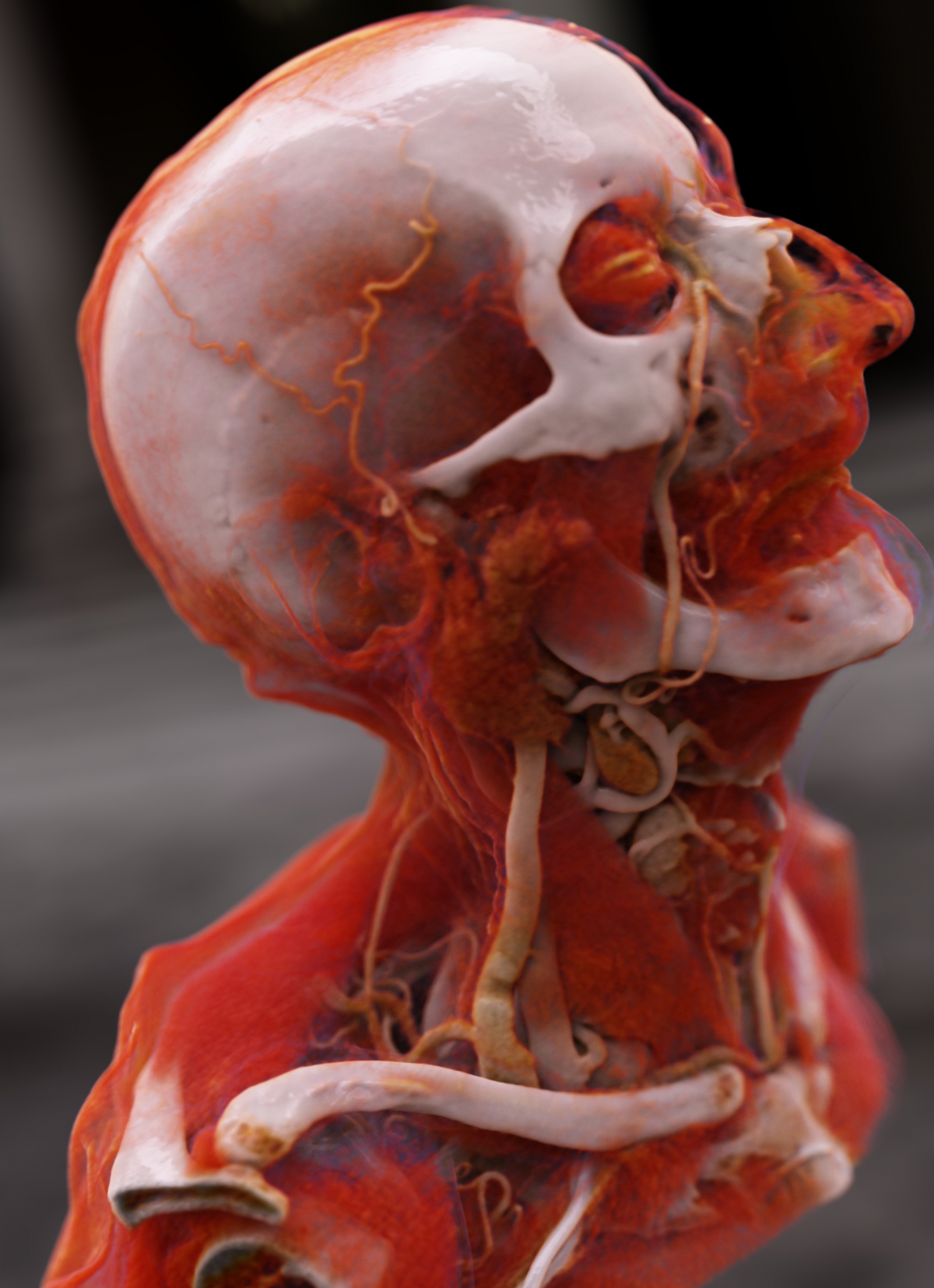
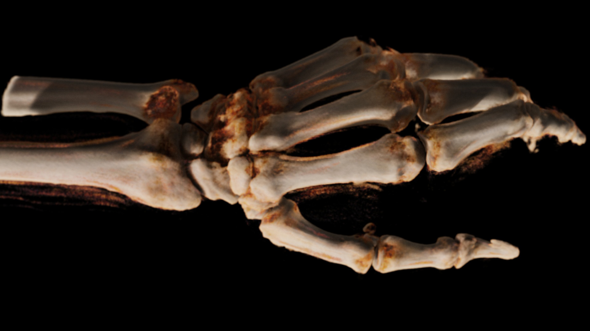
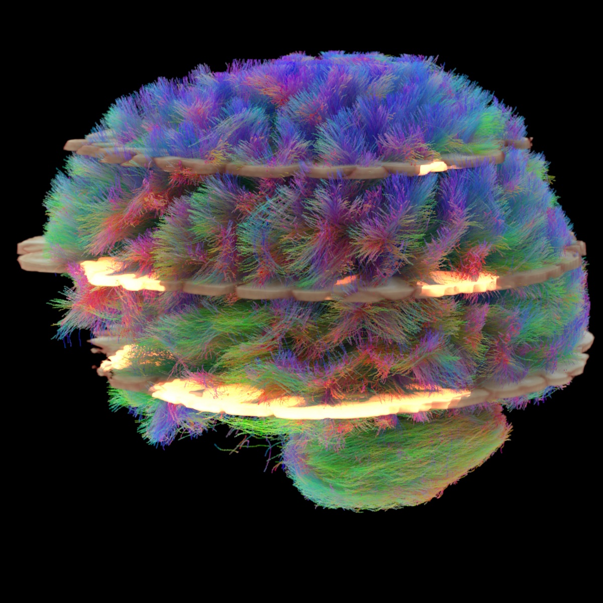
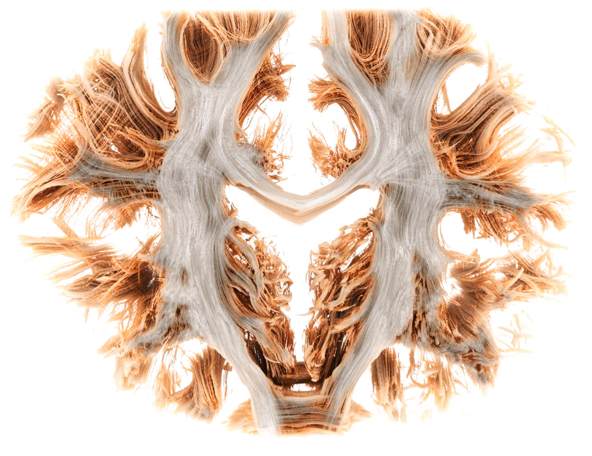
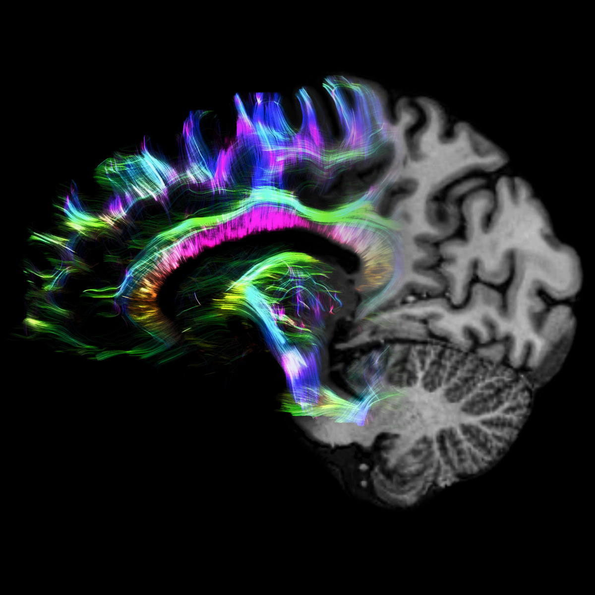
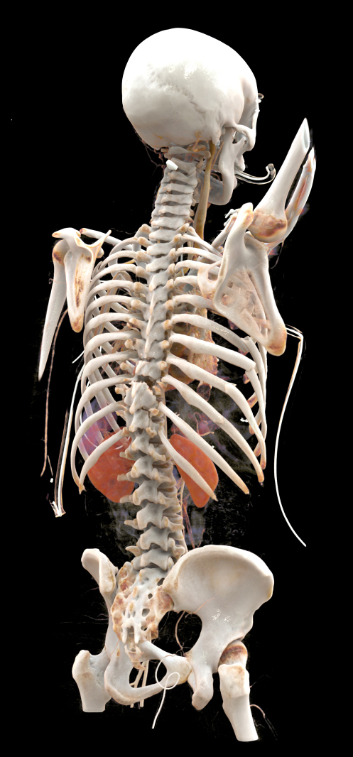
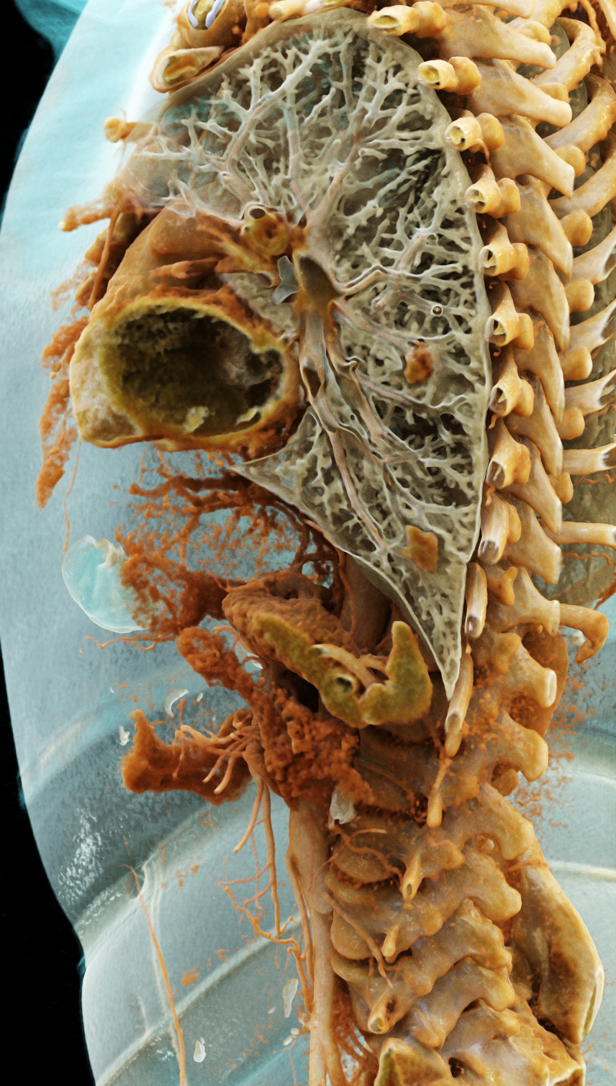
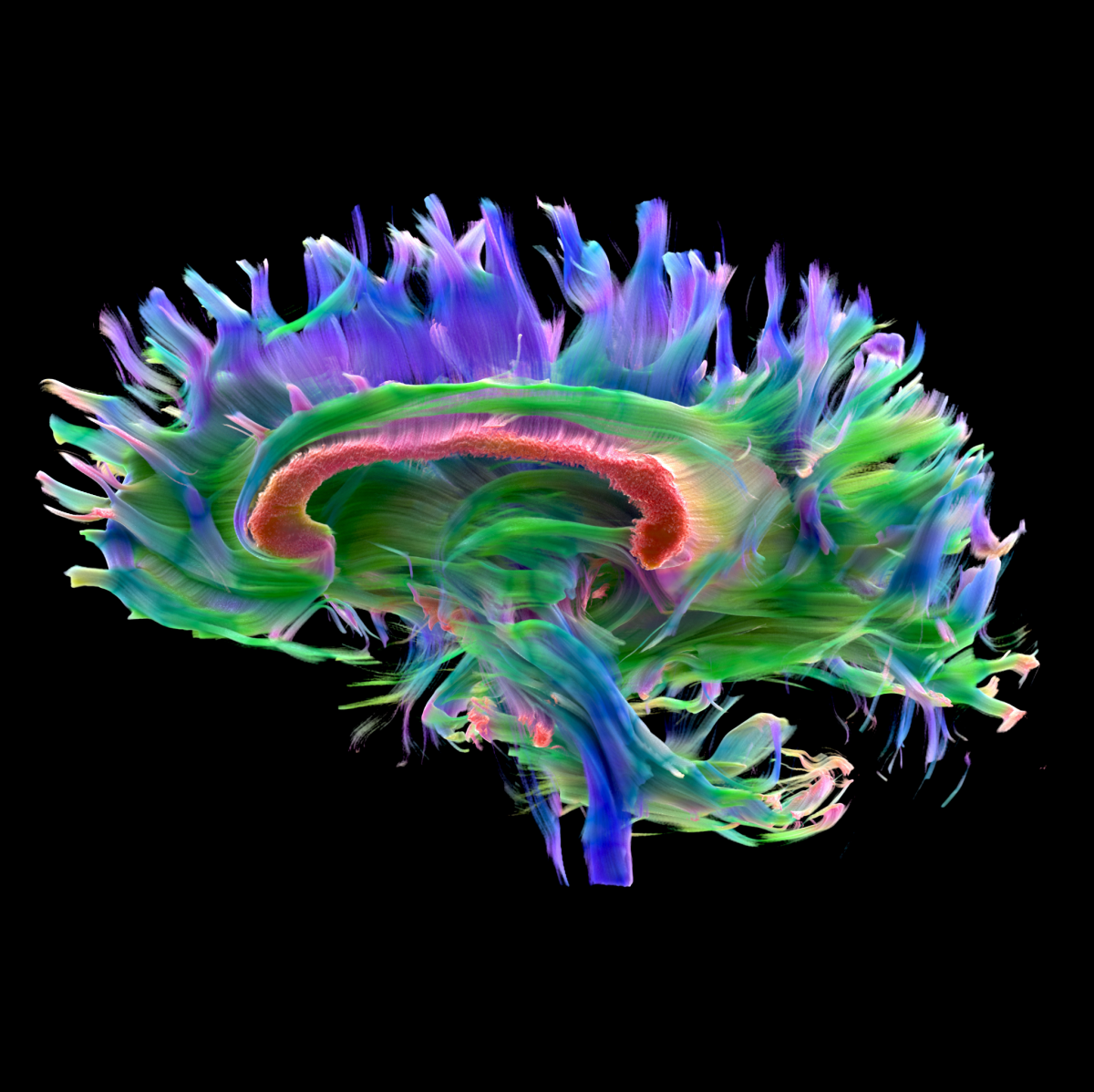
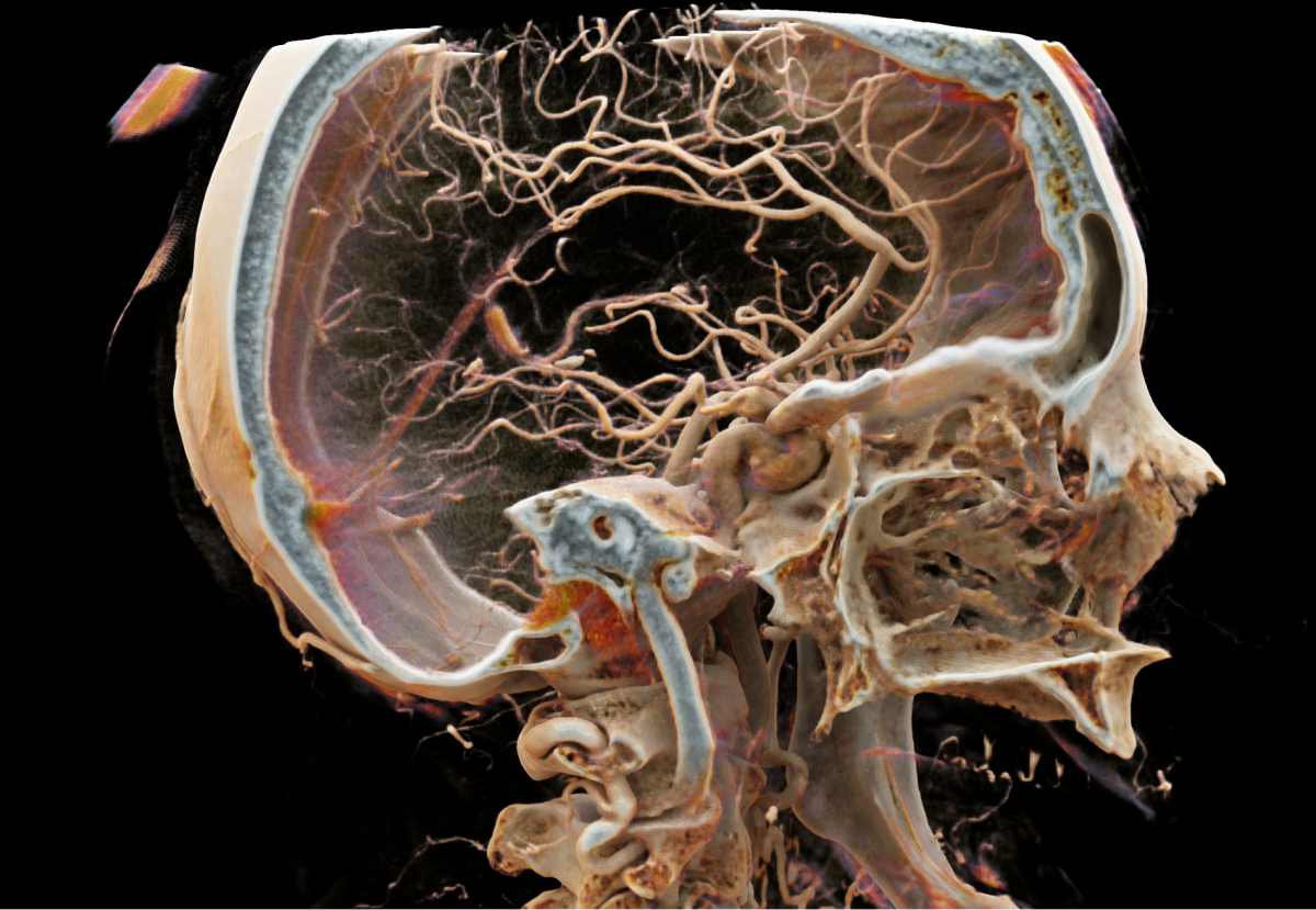
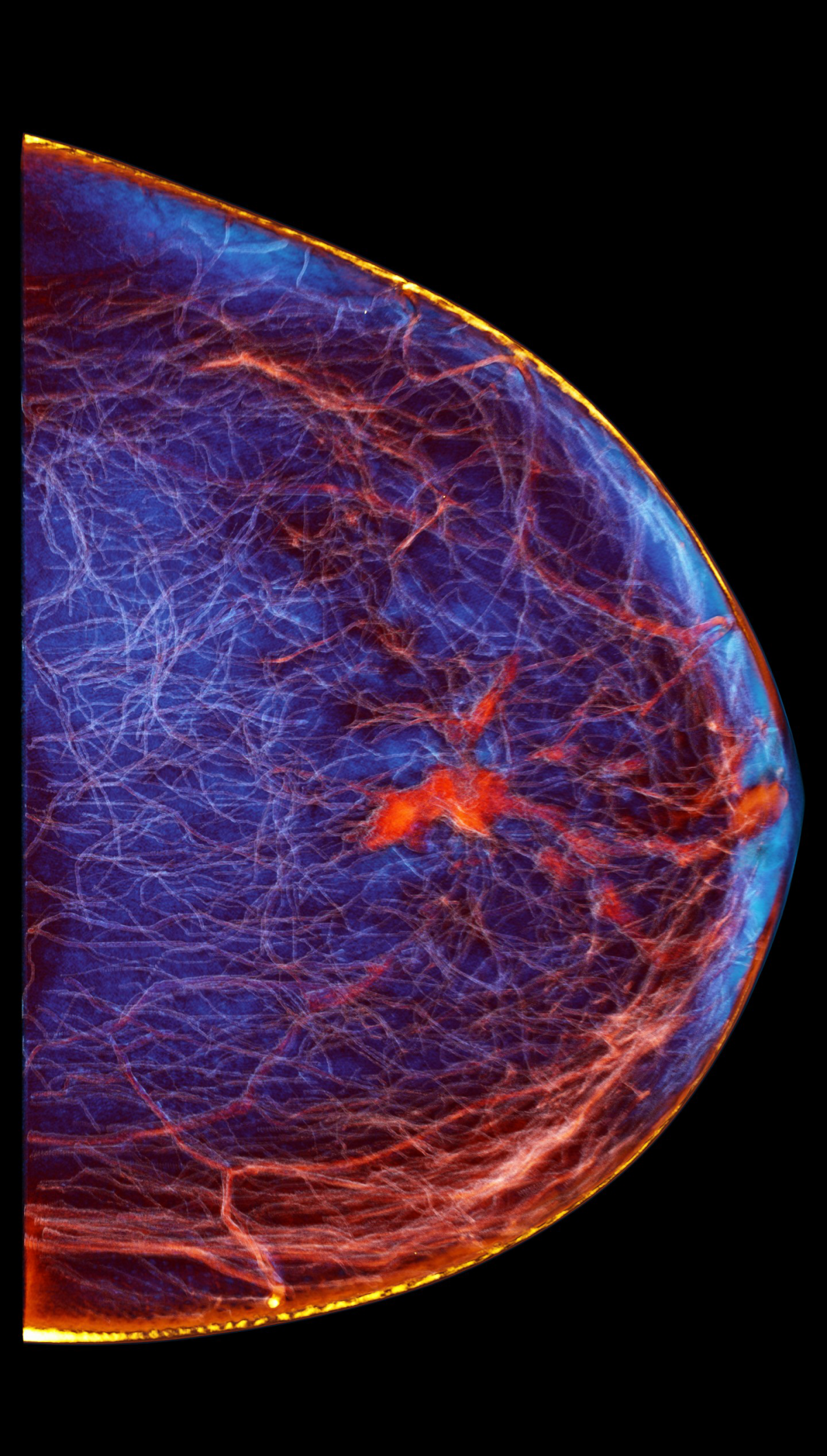
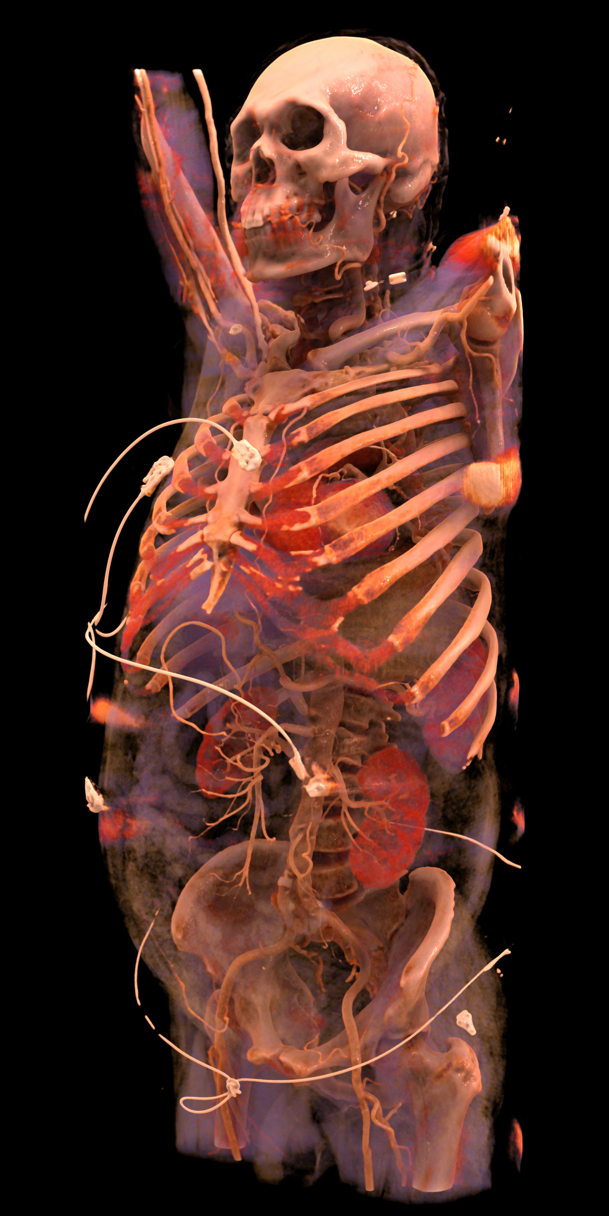
The hyper-realistic computer-generated images often resemble the objects in the “Body worlds” exhibitions. They are based on a sophisticated algorithm which creates spectacular effects, very much like in a Hollywood blockbuster: “Unlike previous 3D imaging procedures, Cinematic Rendering is based on physics,” Dr Engel explains. While before raycasting was used which treats light rays as straight lines, the new technology includes a number of much more complex factors such as reflection and scatter. This results in 3D images in cinematic quality but more importantly with added clinical value. “A crucial feature is realistic shading,” the inventor adds. “The human eye is trained from day 1 to recognize minute differences in shading and deduct the spatial location of an object. A shadow for example can indicate the depth of a given structure. Until today, we did not have this kind of information in 3D imaging which made the evaluation of spatial relationships, such as overlappings, very difficult. The new method offers a more sculptural approach. We consider surgery to be an important field of application where this innovation will be highly useful.”
While Cinematic Rendering is technologically extremely sophisticated, it is versatile in its potential application. “Today, PCs with the necessary computing power are easily available; the same holds true for imaging modalities such as CT, MRI, PET and ultrasound. The system is DICOM-compatible, particular standards are not required,” Dr Engel underlines.

Coming soon to your local OR: augmented reality!
Currently Cinematic Rendering is under development for surgery application. “In the prototype study we focused on therapy planning,” says Dr Stefan Assmann, Director Adjacent Fields at Siemens Healthineers. “The surgeons’ feedback indicates that Cinematic Rendering may help save time.” Professor Grützmann agrees that the new method has enormous potential: “I am sure that many possible applications will surface once the method is being used. Maybe one day we will even be able to show the images in augmented reality during an intervention – with the surgeon wearing special glasses.” Dr Klaus Engel is equally enthusiastic: “Imagine you have pre-op images which you can project onto the patient during the operation. That means you can look inside the patient before the first cut. This would be very useful above all in minimally invasive interventions.”
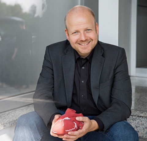
Profile:
Klaus Engel PhD invented the Cinematic Rendering technology. Since 2009, the computer scientist is working as Principal Key Expert at Siemens Healthineers. He specializes in visualization research, prototyping and software development. He spent several years developing and fine-tuning Cinematic Rendering’s algorithms.
25.08.2017


