Image source: OHSU/Christine Torres Hicks
News • Metabolic activity diffusion imaging
New MRI technique reveals cells’ energy activity in organs and tissues
To survive, every cell in the body puts enormous energy into sustaining the right balance of water and essential electrolytes. Researchers at Oregon Health & Science University (OHSU) have developed a way to use magnetic resonance imaging, or MRI, scanning to map this activity in fine detail in the human brain and other organs.
The innovation — called metabolic activity diffusion imaging, or MADI — is opening up new possibilities for detecting cancers and revealing if a tumor is responding to treatment. In upcoming clinical trials enlisting subjects with glioma brain tumors, researchers will compare MADI with positron emission tomography, or PET, which uses injected radioactive agents to create images of cell energy production rates.
Independent experts called it a “compelling mechanistic hypothesis” in an editorial published along with two articles by Springer and co-authors describing MADI in the journal NMR in Biomedicine.

Image source: OHSU/Christine Torres Hicks
“MADI is a new way to make images of metabolic activity within organs and tissues at high spatial resolution, and it’s totally noninvasive,” said inventor Charles Springer, Ph.D., professor with the OHSU Advanced Imaging Research Center. “In principle, this method could apply to almost any pathology. Right now, we are pushing it in the direction of cancer and neuroscience.”
In an animal model using rats, the OHSU researchers already have shown that MADI can detect and monitor brain tumors as effectively as PET, but without the need for injecting tracers or contrast agents of any kind. “It tells us more about what’s going on inside cells in regard to ion transport, water transport, energy production, and so we think it will definitely be useful in cancer and other diseases,” said Martin Pike, Ph.D., associate professor with the OHSU Advanced Imaging Research Center, who is leading the glioma studies.
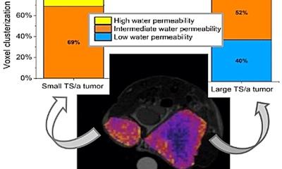
MADI also provides higher resolution images than PET. “It can resolve regions of metabolic activity inside the tumor,” Springer said. “None of the current clinical methods used to map metabolic activity has the spatial resolution needed to measure variations in metabolism within any but the largest tumors.” Ramon Barajas, M.D., associate professor of diagnostic radiology in the OHSU School of Medicine, who is collaborating on the glioma studies, notes that helping to figure out how different parts of a tumor are working can be very useful in making a diagnosis.
MRI works by using a powerful magnetic field to create extremely detailed views of internal organs. The magnetic field causes the nuclei of hydrogen atoms in water molecules to come into alignment with the field. The MRI scanner then delivers radio wave pulses at a resonant frequency. In response, magnetized hydrogen nuclei re-emit radio waves, creating signals that are picked up by the MRI scanner to create images.
MADI builds on a technique called diffusion-weighted MRI, which tracks the movement of water molecules through tissues. Since the 1990s, diffusion-weighted MRI has been used widely in medicine, particularly for brain imaging to detect stroke injury and monitor treatment. The technique delivers fast and informative results without the need for injecting contrast agents. It’s also proving useful for detecting and studying tumors and other disease processes.
It’s like a light bulb, always burning, telling you how much energy the cell is making from the breakdown of the sugar glucose and other nutrients
Charles Springer
But scientists hadn’t fully understood the molecular mechanisms that govern how water molecules move through tissues and cause the changes that become visible signals of stroke and tumors in diffusion MRI. Springer and colleagues pursued the idea that cell membranes play a major role by actively controlling the movement of water molecules in and out of the cells. Their research showed that the probability of water molecules crossing cell membranes is largely dictated by critical enzymes called sodium-potassium pumps. These span cell membranes and pump sodium out and potassium in, a process which also powers the transport of water molecules. “We were able to realize, by learning that water exchange is related to pump activity, that we could make MRI images that map the activity of the sodium-potassium pumps,” Springer said.
The researchers used mathematical modeling and computer simulations to generate information about the movement of water molecules to calculate and map sodium-potassium pump activity. That activity is so crucial to living cells that it serves as a measure of the rate of ongoing energy use. “It’s like a light bulb, always burning, telling you how much energy the cell is making from the breakdown of the sugar glucose and other nutrients,” Springer said. It has never been possible to measure this activity in living things, until now.
Cancer drastically alters energy use in cells, and that is clearly visible in MADI studies using an animal model of glioma brain tumors. “In the animals, we’ve been able to detect cancer, monitor cancer and monitor treatment as well as PET,” Pike said. “We hope we can demonstrate that in humans as well.” Barajas, the neuroradiologist, cautioned that much science remains to be done. “We really have to validate this and make sure that what we're doing is biologically correct.” He said a more detailed and accurate scanning method would greatly benefit patients with brain tumors. “If we get it wrong and stop a therapy that’s actually working, we send them to the operating room for surgery that’s not necessarily needed,” he said. “When we get it wrong and the tumor is growing and we say it’s not, we prevent that patient from getting a new therapy sooner.”
The planned clinical trial aims to recruit about 12 adults with glioma brain tumors. Their brains will be scanned using the PET/MRI hybrid scanner at OHSU — the first in the Pacific Northwest when it was installed in 2021. The hybrid scanner will make it possible to directly compare the performance of PET versus the MADI technique in human subjects.
If MADI proves effective, it could have a number of benefits for patients. The procedure is non-invasive and less time-consuming than PET, taking minutes instead of hours. It’s likely to be less expensive and more widely available than PET. And, MADI can be done using conventional MRI equipment. “PET imaging is the standard of care, but the access for patients is pretty limited to urban areas,” Barajas said. “The ability to do MADI as another standard MRI sequence I think would really open up access to this imaging to a lot more patients that don’t live around urban centers.”
Source: Oregon Health & Science University
04.04.2023