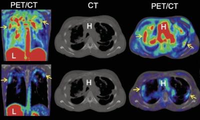
Image courtesy of Prof. He
News • Nuclear medicine
Lung fibrosis: radiopharmaceutical improves diagnosis and monitoring
Trimt GmbH develops novel radiopharmaceuticals for improved detection and treatment of various diseases. While generally focusing on cancer, a recent study linked Trimt's clinical imaging probes to fibrosis, a slowly progressing and lethal disease calling for improved early diagnostics.
Radiopharmaceuticals are radioactive drugs that are used in medical imaging procedures for diagnostics or in radionuclide therapy for targeted tumor treatment. Trimt's proprietary radiopharmaceutical Ga-68-Trivehexin is a so-called radiotracer for positron emission tomography (PET). At Zhongnan Hospital in Wuhan (China)1 and Istanbul University in Turkey2, Ga-68-Trivehexin was recently used to accurately diagnose patients with pulmonary fibrosis and concomitant lung cancer using positron emission tomography (PET).
The amazing imaging of lung fibrosis with Ga-68-Trivehexin indicates that this radiotracer offers a transformative approach to non-invasively visualizing αvβ6-integrin expression, a key driver of TGF-β-mediated fibrosis
Yong He
Lung fibrosis (especially idiopathic pulmonary fibrosis, IPF) is a disease characterized by scarring and thickening of lung tissue. This scarring makes the lungs stiff and impairs the breathing ability. Patients often suffer from dry cough and fatigue. As disease progresses, the need for oxygen therapy reduces their independence and ability to work, and their quality of life is often reduced by depression and anxiety. Lung fibrosis also significantly increases the risk of developing lung carcinoma, the most prevalent form of lung cancer.
Early diagnosis would allow for timely initiation of anti-fibrotic medication, which can slow the decline in lung function and maintain patients' life quality. Treatment monitoring is crucial during therapy for pulmonary fibrosis. Reliable assessment of therapeutic efficacy and/or early detection of disease progression would enable to adjust treatment plans timely, such as switching to more aggressive interventions if necessary, and thus, improve life expectancy.
Currently, identification of IPF in its early stages is challenging due to non-specific symptoms and difficult differential diagnosis. Non-invasive radiological procedures such as computed tomography (CT) images often delivers results suggestive of IPF but not definitive. CT diagnosis may be confirmed by invasive lung biopsy, which however carries additional risks and may not be recommended for all patients. The complexity of IPF detection underscores the imperative to refine diagnostic imaging methods.

Photo kindly provided by Prof He
Yong He, Director of Nuclear Medicine at Zhongnan Hospital, Wuhan University, led the team that reported the first application of Ga-68-Trivehexin for PET/CT imaging of IPF in early March of 2025.1 "The amazing imaging of lung fibrosis with Ga-68-Trivehexin indicates that this radiotracer offers a transformative approach to non-invasively visualizing αvβ6-integrin expression, a key driver of TGF-β-mediated fibrosis", he commented the paper. "It enables precise mapping of active fibrotic regions in the lung. Furthermore, it may allow clinicians to monitor fibrotic activity in real time, assess treatment response for in αvβ6-integrin targeted interventions, including anti-αvβ6-monoclonal antibody and small molecular inhibitors currently under investigation in clinical trials".
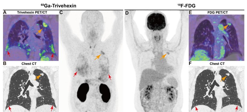
Only a few days later, a similar case was published by Serkan Kuyumcu and collegues from Department of Nuclear Medicine, Istanbul University.2 The authors pointed out that integrin imaging may provide insights into the dynamic processes of fibrosis, bridging diagnostic and therapeutic applications. They concluded that Ga-68-Trivehexin uptake in fibrosis offers a promising avenue for monitoring fibrotic diseases, warranting further research to optimize the clinical utility of integrin-targeted PET imaging.
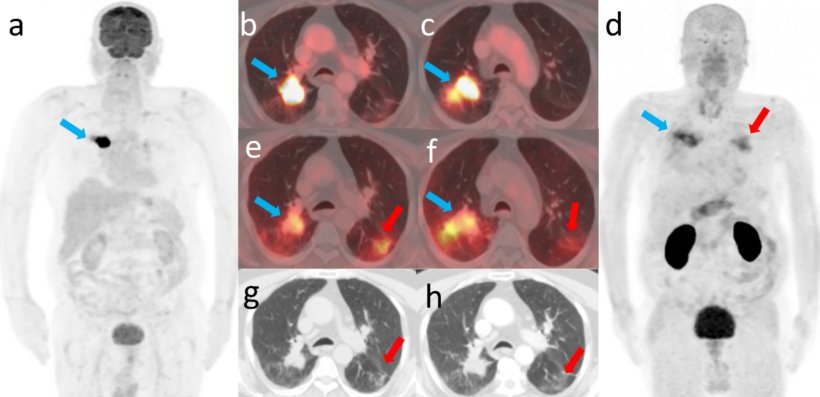
Image source: Kuyumcu S, Denizmen Zorba D, Özkan ZG, European Journal of Nuclear Medicine and Molecular Imaging 2025 (CC BY 4.0)
Ga-68-Trivehexin is based on a biomolecule (more precisely: a peptide) that selectively binds to a cell surface protein called αvβ6-integrin. The radiotracer was originally developed by Johannes Notni and coworkers at Technical University of Munich, who is now the Chief Science Officer of Trimt. "Ga-68-Trivehexin enabled an astonishingly high-contrast visualization of the fibrotic lung tissue in the PET image. Beyond that, one of the PET scans also revealed the presence of a rare form of lung cancer which presumably had developed in the course of fibrosis progression. These examples demonstrate that assessment of the complex disease patterns of IPF might be facilitated by Ga-68-Trivehexin PET/CT in a one-stop-shop fashion, and furthermore could be useful for follow-up during therapy", said Notni, describing the implications of the study.
"The high relevance of these results is underscored by the fact that the standard PET tracer F-18-FDG, a radioactive sugar molecule, failed to detect the fibrosis in thes cases", said Jakub Šimeček, CEO of Trimt. "We congratulate the clinical investigators, Prof. Yong He, Prof. Serkan Kuyumcu, and their teams, to highlighting the potential added value of Ga-68-Trivehexin PET imaging in such a clinically challenging scenario". These results were recently published in the European Journal of Nuclear Medicine and Medical Imaging, one of the most influential journals in the field, and highlighted as "Image of the Month".
Scientific publications:
- Huiqin Wu, Ling Li, Zhiwei Xiao, Qiongrong Chen, Chongjiao Li, Yong He. [68Ga]Ga-Trivehexin PET/CT imaging of integrin-αvβ6 expression in concomitant mucinous lung adenocarcinoma and idiopathic pulmonary fibrosis. Eur J Nucl Med Mol Imaging (2025).
- Serkan Kuyumcu, Dilara Denizmen Zorba, Zeynep Gözde Özkan. 68Ga-Trivehexin PET/CT uptake in malignant and fibrotic lung tissue: refining diagnostic applications. Eur J Nucl Med Mol Imaging (2025).
Source: Trimt GmbH
24.03.2025