Interview • Hybrid imaging
‘We need to drive standardisation in scanning’
During our interview with Professor Regina Beets-Tan, President of the European Society of Radiology (ESR), we asked about possible future developments in cancer imaging.
Interview: Michael Krassnitzer
Image source: Shutterstock/SvedOliver
Beets-Tan unhesitatingly pointed out that screening exams are ever-increasing and thus more and more tumours are and will be detected at an earlier stage. ‘The earlier a tumour is detected,’ she added, ‘the better it can be treated: by resection, minimally invasive surgery, or by interventional radiology. This means we need very sensitive imaging techniques that can identify very small tumours, and also tumour growth. So we need high-resolution anatomical imaging combined with biological imaging that visualises how the tumour is behaving.
‘Treatment of malignancies where the tumour has already spread to other parts of the body has also become more effective – the keywords are immunotherapy and targeted therapy. To evaluate the response of these therapies, we also need special imaging techniques that can visualise not only the morphology, but also the biology of the tumour. In addition, we want to extract as much information as possible from these images by using artificial intelligence (AI).’
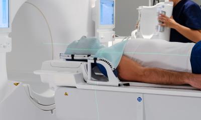
‘Hybrid imaging. That's the future. It involves the combination of several techniques: First, magnetic resonance imaging (MRI), which provides high-resolution images of morphology; second, perfusion imaging, which can quantify blood flow to the tumour; third, diffusion imaging that can map the cellular architecture, which is quite different in cancer cells than in healthy cells; and fourth, positron emission tomography (PET), which can map the metabolic activity of a tumour. Combining all these methods gives the highest possible sensitivity.
‘Currently, imaging capabilities in immunotherapy are very limited. Computed tomography (CT) is used in follow-up scans, but this morphologic imaging alone is insufficient. Sometimes the tumour becomes larger during treatment, but this is not tumour growth but swelling due to the immune response. Therefore, biopsies have to be done regularly to find out what the response of the tumour to immunotherapy actually looks like. In other words, we also need imaging here to visualise the behaviour of the tumour. Specifically, we need to visualise the tumour microenvironment, that is, the immediate environment of the tumour, down to the level of the individual immune cell.’
How far developed is such imaging?
‘We are still at the very beginning. First, it’s important to get funding for the relevant research. I’m a member of the Mission Board for cancer, an expert body that advises the EU Commission on cancer. We have recommended that the Commission invest in research into modern hybrid technologies to better evaluate the response to the new, very expensive therapies - such as immunotherapy.’
Has the initial euphoria faded about AI’s potential to reveal tumours?
Once the underlying data is standardised, the results obtained using AI will also be more reliable
Regina Beets-Tan
‘AI is still a pretty promising thing. However, it has proven to be a problem that the data with which the artificial intelligence is fed must be very homogeneous. At my clinic, a study has just been completed in which 800 MRI images of a particular tumour from all over the Netherlands have been fingerprinted by an AI. The results are little better than a coin toss. That's precisely because the scans are all so different. So we need to drive standardisation. Once the underlying data is standardised, the results obtained using AI will also be more reliable.’
Alongside future cancer imaging, what might arrive to advance radiology in general?
‘Radiologists will play a greater role in the clinic in the future. Currently, radiologists are busy with a lot of routine tasks, which in the future can be taken over by AI, and also by less specialised and less expensive staff. The time radiologists gain when relieved of routine tasks can be put into other tasks. Why shouldn't the radiologist explain his/her image findings to patients?
‘Now that malignant diseases are being detected earlier and earlier, the need for interventional radiology will also increase. In any case, radiology must continue to evolve. We need to combine our technology with clinical expertise. If we don't, we run the risk that our specialty could one day become superfluous.’
Profile:
Professor Regina Beets-Tan is President of the European Society of Radiology (ESR) and Chair Department of Radiology at the Netherlands Cancer Institute, Amsterdam.
28.11.2021