Article • Innovations in imaging
Latest advances in CT, MRI & PET might not enter clinic soon
Current advances such as phase-contrast CT are taking medical imaging further but their use in clinical practice may have to wait up to a decade, prominent physicist predicts.
Report: Mélisande Rouger
Cooperation between medical specialists and biomedical engineers is having a fresh start as healthcare professionals were invited to the annual meeting of the Spanish Biomedical Engineers Society (CASEIB 2016) in November in Valencia. Researchers and key representatives from all across the country shared expertise during dedicated roundtables and sessions to try and tailor technological advances to current healthcare needs, notably in medical imaging. During the meeting, José María Benlloch, a professor of investigation at the Spanish Superior Council for Scientific Investigation at Valencia Polytechnic University, presented the newest advances in medical imaging techniques. The long held perception that CT is better for hard tissues imaging and MRI better for soft tissues is about to become history, according to Benlloch, who obtained the Spanish National Investigation Award in 2014 for applying particle physics techniques to molecular imaging in biomedicine. However, years will pass before some of these improvements can benefit population healthcare, he believes. ‘Phase-contrast will change the paradigm and bring CT into soft tissue imaging. However the technique will not enter clinical practice before 10 to 15 years, because we first need a coherent source of X-rays. I believe X-ray techniques using laser will become such a source and will be much more affordable in the future.’
Unlike CT spectroscopy, a mature technique that will enter clinical practice in three to five years according to Benlloch, phase-contrast CT is still facing a number of technological challenges. For X-ray detectors to be sensitive to phase-contrast, resolutions in the one micrometer range are needed. Researchers are working on these developments, but they still have a long road ahead.’ We can achieve this resolution by placing a rack on detectors with poorer resolution, but in doing so we lose a lot of X-ray power. Clearly significant technological advances are still lacking for the technique to become financially available and be used in most hospitals,’ he said.
MRI’s trademark difficulty in imaging hard tissues could soon be solved as well, as Benlloch and his team are currently working on MRI solutions for dental applications, in which dentists could not only see teeth but also the surrounding soft tissues. ‘This is very interesting for dentists because it will not use radiation. They will be able to image gums and things that are now almost invisible on X-ray,’ he said. This new MRI development uses a permanent magnet applied to only a small part of the body. It also uses a low electromagnetic field and does not occupy as much space as current scanners. Acquisition time will be just a minute and the system will be automatised so that dentists can read the scans themselves without the help of an MR specialist. The cost will also be significantly inferior to traditional MR scanners - €120,000, experts estimate. The technique should be ready by the end of 2017, and available in clinic in two to three years if it receives FDA and CE marking.
It is better that radiologists participate from the start so that we don’t lose time building a machine they may not need
Jose Maria Benlloch
In PET imaging, the US National Institute of Health has recently granted $15 million to California University UC Davis, Berkeley University and Pennsylvania University to develop a true whole-body PET/CT scanner enabling to image the patient in 2 minutes while significantly reducing dose. ‘This will improve sensitivity in diseases and tumors detection and enable to acquire dynamic pictures of the whole body. We will be able to see biology as never before, for instance we will be able to see how molecules evolve from one part of the body to another and in how much time, we will have temporal resolution. Being able to see these correlations in real time will bring much more information than what we currently have,’ Benlloch said. The fact that radiologists are coming to the congress will help engineers better understand the needs of their colleagues, the researcher believes. ‘It is better that radiologists participate from the start so that we don’t lose time building a machine they may not need.’ ‘Cooperation between doctors and engineers is absolutely necessary for the investigation and the development of new technologies that improve citizens quality of life. The doctors in their daily clinical practice may detect the limitations of current technology and the problems to be solved, and from that information the engineers can offer solutions and improvements. To be efficient, this process needs a very tight cooperation between both parties, and the relatively recent introduction of the biomedical engineering specialty in Spain will impulse in this direction,’ Francisco Javier Saiz Rodriguez, president of CASEIB 2016 organising committee, echoed.
The Spanish Society of Radiology (SERAM) will also welcome biomedical engineers during its next biannual meeting in 2018, as the congress scientific committee will dedicate an area to biomedical engineering, making it an important line of the meeting. ‘For us this strategic positioning. There are many on-going cooperation projects between the two specialties in Spain, but we don’t have a registry yet. We are still very much unaware of what is being done and what could be done with biomedical engineering. There are many areas of cooperation that we could work on, such as Big Data use, standardisation of new techniques and image acquisition itself – for instance to obtain an image of the heart without performing apnea in some cardiac settings. I think our cooperation is just taking off,’ Àngel Gayete, SERAM President, said.
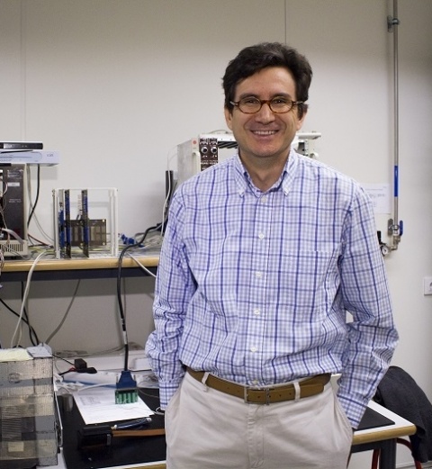
Profile:
Jose Maria Benlloch is a professor of investigation at the Spanish Superior Council for Scientific Investigation (CISC) at Valencia Polytechnic University and Director of the Instrumentation Institute for Molecular Imaging at CISC. He received the National Investigation Prize in 2014 for his relevant contributions to the application of particle physics techniques to molecular imaging in biomedicine. Benlloch wrote his PhD thesis in 1990 on detector development and data analysis in the DELPHI experiment of the LEP collider at CERN in Geneva, Switzerland. He has been on the research staff at the Massachusetts Institute of Technology under the supervision of Physics Nobel Prize holder Jerôme Friedman and has published over 200 articles in international publications. He has coordinated 30 research projects and has eight patents and three spin-off companies in biomedical engineering.
16.06.2017