Article • From technology to responsibility
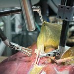
AI in surgery – tool, or surgeon of tomorrow?
Will surgeons be replaced by machines in the future? With the rising impact of AI and robotics, this concern is on the minds of many medical professionals and patients alike. At the 2026 German Surgery Congress, Prof. Dr Jörg-Peter Ritz, President of the German Society for General and Visceral Surgery (DGAV) and Chief Physician and Medical Director at Helios Kliniken Schwerin, painted a more…