Image credit: EPFL/.Neurorestore (CC BY-SA 4.0)
News • Study shows potential to reverse paralysis
Another step towards regeneration after spinal cord injuries
When the spinal cords of mice and humans are partially damaged, the initial paralysis is followed by the extensive, spontaneous recovery of motor function. However, after a complete spinal cord injury, this natural repair of the spinal cord doesn’t occur and there is no recovery.
Meaningful recovery after severe injuries requires strategies that promote the regeneration of nerve fibers, but the requisite conditions for these strategies to successfully restore motor function have remained elusive.
“Five years ago, we demonstrated that nerve fibers can be regenerated across anatomically complete spinal cord injuries,” says Mark Anderson, a senior author of the study. “But we also realized this wasn’t enough to restore motor function, as the new fibers failed to connect to the right places on the other side of the lesion.” Anderson is the director of Central Nervous System Regeneration at .NeuroRestore and a scientist at the Wyss Center for Bio and Neuroengineering.
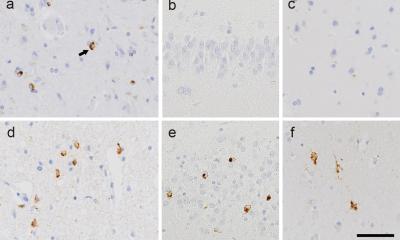
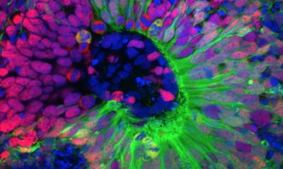
Working in tandem with peers at UCLA and Harvard Medical School, the scientists used state-of-the-art equipment at EPFL’s Campus Biotech facilities in Geneva to run in-depth analyses and identity which type of neuron is involved in natural spinal-cord repair after partial spinal cord injury. “Our observations using single-cell nuclear RNA sequencing not only exposed the specific axons that must regenerate, but also revealed that these axons must reconnect to their natural targets to restore motor function,” says Jordan Squair, the study’s first author. The team’s findings appear in Science.
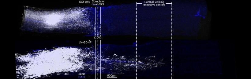
Image credit: EPFL/.Neurorestore (CC BY-SA 4.0)
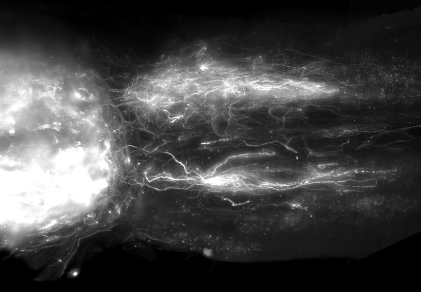
Their discovery informed the design of a multipronged gene therapy. The scientists activated growth programs in the identified neurons in mice to regenerate their nerve fibers, upregulated specific proteins to support the neurons’ growth through the lesion core, and administered guidance molecules to attract the regenerating nerve fibers to their natural targets below the injury. “We were inspired by nature when we designed a therapeutic strategy that replicates the spinal-cord repair mechanisms occurring spontaneously after partial injuries,” says Squair.
We believe a complete solution for treating spinal cord injury will require both approaches – gene therapy to regrow relevant nerve fibers, and spinal stimulation to maximize the ability of both these fibers and the spinal cord below the injury
Grégoire Courtine
Mice with anatomically complete spinal cord injuries regained the ability to walk, exhibiting gait patterns that resembled those quantified in mice that resumed walking naturally after partial injuries. This observation revealed a previously unknown condition for regenerative therapies to be successful in restoring motor function after neurotrauma. “We expect that our gene therapy will act synergistically with our other procedures involving electrical stimulation of the spinal cord,” says Grégoire Courtine, a senior author of the study who also heads .NeuroRestore together with Jocelyne Bloch. “We believe a complete solution for treating spinal cord injury will require both approaches – gene therapy to regrow relevant nerve fibers, and spinal stimulation to maximize the ability of both these fibers and the spinal cord below the injury to produce movement.”
While many obstacles must still be overcome before this gene therapy can be applied in humans, the scientists have taken the first steps towards developing the technology necessary to achieve this feat in the years to come.
Source: EPFL
22.09.2023