Image source: Baadsvik EL et al., Magnetic Resonance in Medicine 2024 (CC BY-NC 4.0)
News • Myelin visualisation
New MRI procedure makes multiple sclerosis visible
ETH Zurich researchers have developed a new method of magnetic resonance imaging (MRI) for the early detection and better monitoring of multiple sclerosis (MS).
Multiple sclerosis is a neurological disease that usually leads to permanent disabilities. It affects around 2.9 million people worldwide, and around 15,000 in Switzerland alone. One key feature of the disease is that it causes the patient’s own immune system to attack and destroy the myelin sheaths in the central nervous system. These protective sheaths insulate the nerve fibres, much like the plastic coating around a copper wire. Myelin sheaths ensure that electrical impulses travel quickly and efficiently from nerve cell to nerve cell. If they are damaged or become thinner, this can lead to irreversible visual, speech and coordination disorders.
So far, however, it hasn’t been possible to visualise the myelin sheaths well enough to use this information for the diagnosis and monitoring of MS. Now researchers at ETH Zurich and University of Zurich, led by Markus Weiger and Emily Baadsvik from the Institute for Biomedical Engineering, have developed a new MRI procedure that maps the condition of the myelin sheaths more accurately than was previously possible. The researchers successfully tested the procedure on healthy people for the first time.
Their insights have been published in the journals Magnetic Resonance in Medicine and Science Advances.
In the future, the MRI system with its special head scanner could help doctors to recognise MS at an early stage and better monitor the progression of the disease. The technology could also facilitate the development of new drugs for MS. But it doesn’t end there: the new MRI method could also be used by researchers to better visualise other solid tissue types such as connective tissue, tendons and ligaments.
Conventional MRI devices capture only inaccurate, indirect images of the myelin sheaths. That’s because most of these devices work by reacting to water molecules in the body that have been stimulated by radio waves in a strong magnetic field. But the myelin sheaths, which wrap around the nerve fibres in several layers, consist mainly of fatty tissue and proteins. That said, there is some water – known as myelin water – trapped between these layers. Standard MRIs build their images primarily using the signals of the hydrogen atoms in this myelin water, rather than imaging the myelin sheaths directly.
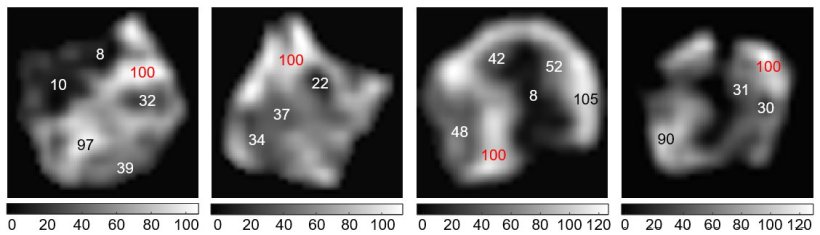
The ETH researchers’ new MRI method solves this problem and measures the myelin content directly. It puts numerical values on MRI images of the brain to show how much myelin is present in a particular area compared to other areas of the image. A number 8, for instance, means that the myelin content at this point is only 8 percent of a maximum value of 100, which indicates a significant thinning of the myelin sheaths. Essentially, the darker the area and the smaller the number in the image, the more the myelin sheaths have been reduced. This information ought to enable doctors to better assess the severity and progression of MS.
However, it is difficult to image the myelin sheaths directly. That’s because the signals that the MRI triggers in the tissue are very short-lived; the signals that emanate from the myelin water last much longer. “Put simply, the hydrogen atoms in myelin tissue move less freely than those in myelin water. That means they generate much briefer signals, which disappear again after a few microseconds,” Weiger says, adding: “And bearing in mind a microsecond is a millionth of a second, that’s a very short time indeed.” A conventional MRI scanner can’t capture these fleeting signals because it doesn’t take the measurements fast enough.
To solve this problem, the researchers used a specially customised MRI head scanner that they have developed over the past ten years together with the companies Philips and Futura. This scanner is characterised by a particularly strong gradient in the magnetic field. “The greater the change in magnetic field strength generated by the three scanner coils, the faster information about the position of hydrogen atoms can be recorded,” Baadsvik says.

Image source: ETH Zurich
Generating such a strong gradient calls for a strong current and a sophisticated design. As the researchers scan only the head, the magnetic field is more contained and concentrated than with conventional devices. In addition, the system can quickly switch from transmitting radio waves to receiving signals; the researchers and their industry partners have developed a special circuit for this purpose.
The researchers have already successfully tested their MRI procedure on tissue samples from MS patients and on two healthy individuals. Next, they want to test it on MS patients themselves. Whether the new MRI head scanner will make its way into hospitals in the future now depends on the medical industry. “We’ve shown that our process works,” Weiger says. “Now it’s up to industry partners to implement it and bring it to market.”
Source: ETH Zurich; author: Christoph Elhardt
10.02.2024