News • Diffusion-based generative model
Better MRI scans of beating hearts with AI
Model generates higher quality scans in less time, improves patient experience
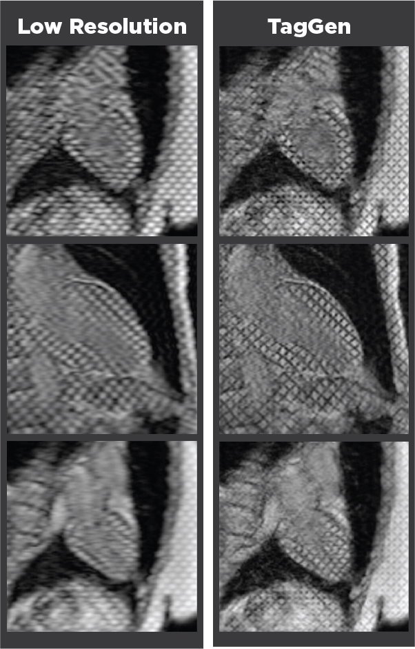
Image credit: MU Health Care, Claudia Carver
An AI-assisted model developed by researchers from the University of Missouri (Mizzou) School of Medicine and the School of Engineering can take low-quality MRI heart scans and turn them into high quality images, while reducing the time needed to scan the heart by about 90%.
The researchers present the model in the journal Magnetic Resonance in Medicine.
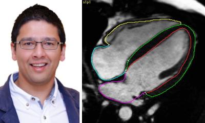
A cardiac magnetic resonance imaging (MRI) scan usually takes anywhere from 30 to 90 minutes. The scans can reveal important information on how well the heart is working and if there are any problems. But they aren’t always clear or of great quality, as movement can reduce image quality. That’s where TagGen, the AI-assisted model developed by Mizzou researchers, can make a big difference. “If you have a blurry image, you have very few ways to recover the fine details or quality of the image,” said Changyu Sun, the lead researcher. “The sharpness reveals very important information for the clinical diagnosis, like if there’s abnormal movement or any dysfunction.”
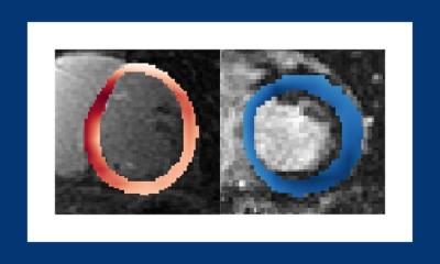
The better quality means the scans have sharper taglines, which are markers that track muscle movement. These taglines help doctors identify areas of the heart that aren’t moving properly or may be damaged. Without them, it can be difficult to track the motion or accurately measure cardiac function. As the AI processes the image, it restores the quality and provides better visualization of the heart moving.
By using TagGen to maintain the taglines, doctors can see information they would have otherwise missed, and patients only need to hold their breath for three heartbeats
Changyu Sun
The faster scanning and improved tagline quality enabled by AI allows doctors to better observe the heart and how it beats, contracts and pumps. Without TagGen, acquiring the scan would take significantly longer and would drive up cost, patient discomfort and lower image quality.
"During a heart MRI scan, patients are asked to hold their breath to reduce chest movement from breathing, which helps create clearer images.” Sun said. “Some scans take more than 20 heartbeats, making it harder for patients to hold their breath. By using TagGen to maintain the taglines, doctors can see information they would have otherwise missed, and patients only need to hold their breath for three heartbeats. This technology will lead to better diagnoses and improved patient outcomes.”
For future work, Sun plans on refining TagGen and improving the MRI motion tracking. He and his team are also working on generalizing the AI technique to other types of cardiac MRI scans, computed tomography (CT) scans and scans for other organs, like a brain MRI.
Source: University of Missouri-Columbia
29.06.2025