News • Ultraviolet cell visualisation
'Seeing' cancer though the eyes of a butterfly
There are many creatures on our planet with more advanced senses than humans. Turtles can sense Earth’s magnetic field. Mantis shrimp can detect polarized light. Elephants can hear much lower frequencies than humans can. Butterflies can perceive a broader range of colors, including ultraviolet (UV) light.
Image credit: The Grainger College of Engineering at University of Illinois Urbana-Champaign
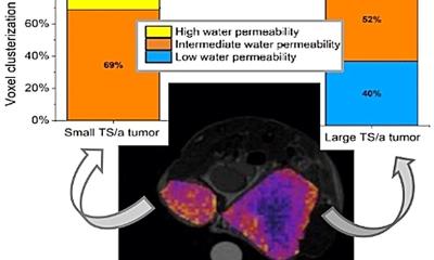
Inspired by the enhanced visual system of the Papilio xuthus butterfly, a team of researchers have developed an imaging sensor capable of “seeing” into the UV range inaccessible to human eyes. The design of the sensor uses stacked photodiodes and perovskite nanocrystals (PNCs) capable of imaging different wavelengths in the UV range. Using the spectral signatures of biomedical markers, such as amino acids, this new imaging technology is even capable of differentiating between cancer cells and normal cells with 99% confidence.
This new research, led by University of Illinois Urbana-Champaign electrical and computer engineering professor Viktor Gruev and bioengineering professor Shuming Nie, was recently published in the journal Science Advances. Both Gruev and Nie are affiliates of the Cancer Center at Illinois.
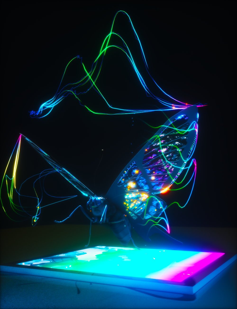
Image credit: The Grainger College of Engineering at University of Illinois Urbana-Champaign
“We've taken inspiration from the visual system of butterflies, who are able to perceive multiple regions in the UV spectrum, and designed a camera that replicates that functionality,” Gruev says. “We did this by using novel perovskite nanocrystals, combined with silicon imaging technology, and this new camera technology can detect multiple UV regions.”
UV light is electromagnetic radiation with wavelengths shorter than that of visible light (but longer than x-rays). We are most familiar with UV radiation from the sun and the dangers it poses to human health. UV light is categorized into three different regions—UVA, UVB and UVC— based on different wavelength ranges. Because humans cannot see UV light, it is challenging to capture UV information, especially discerning the small differences between each region.
Butterflies, however, can see these small variations in the UV spectrum, like humans can see shades of blue and green. Gruev notes, “It is intriguing to me how they are able to see those small variations. UV light is incredibly difficult to capture, it just gets absorbed by everything, and butterflies have managed to do it extremely well.”
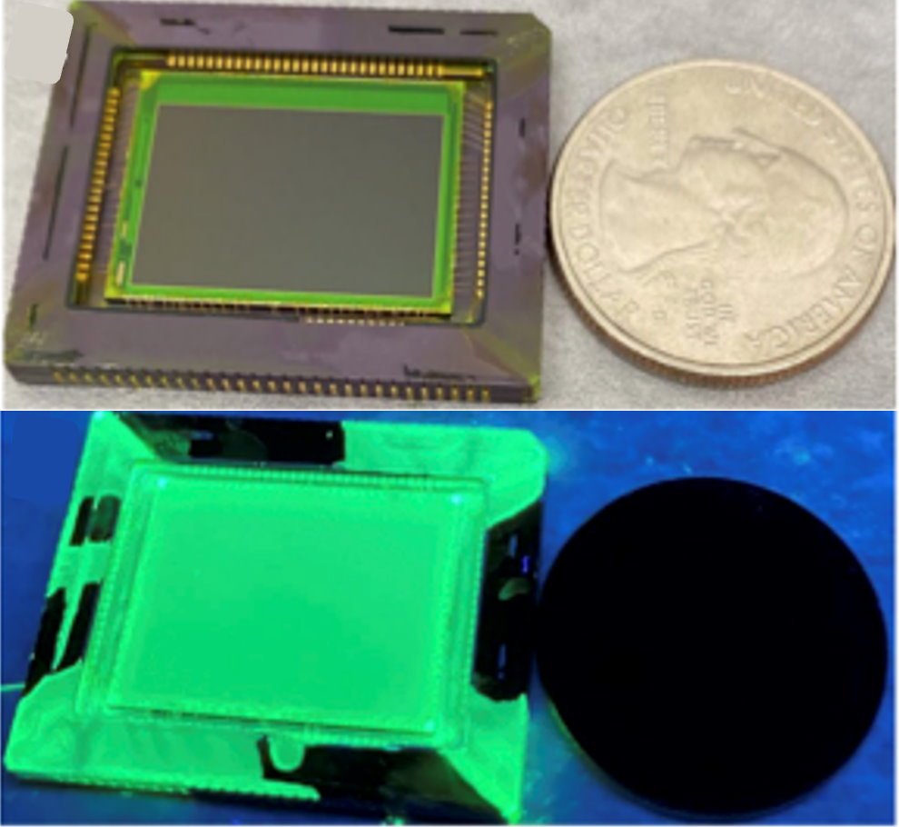
Image source: The Grainger College of Engineering at University of Illinois Urbana-Champaign; adapted from: Chen et al., Science Advances 2023 (CC BY 4.0)
Humans have trichromatic vision with three photoreceptors, where every color perceived can be made from a combination of red, green and blue. Butterflies, however, have compound eyes, with six (or more) photoreceptor classes with distinct spectral sensitivities. In particular, the Papilio xuthus, a yellow, Asian swallowtail butterfly, has not only blue, green and red, but also violet, ultraviolet and broadband receptors. Further, butterflies have fluorescent pigments that allow them to convert UV light into visible light which can then be easily sensed by their photoreceptors. This allows them to perceive a broader range of colors and details in their environment.
Beyond the increased number of photoreceptors, butterflies also exhibit a unique tiered structure in their photoreceptors. To replicate the UV sensing mechanism of the Papilio xuthus butterfly, the UIUC team has emulated the process by combining a thin layer of PNCs with a tiered array of silicon photodiodes.
PNCs are a class of semiconductor nanocrystals that display unique properties similar to that of quantum dots—changing the size and composition of the particle changes the absorption and emission properties of the material. In the last few years, PNCs have emerged as an interesting material for different sensing applications, such as solar cells and LEDs. PNCs are extremely good at detecting UV (and even lower) wavelengths that traditional silicon detectors are not. In the new imaging sensor, the PNC layer is able to absorb UV photons and re-emit light in the visible (green) spectrum which is then detected by the tiered silicon photodiodes. Processing of these signals allows for mapping and identification of UV signatures.
Image source: The Grainger College of Engineering at University of Illinois Urbana-Champaign
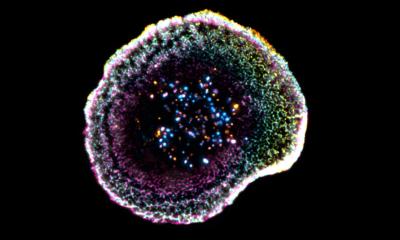
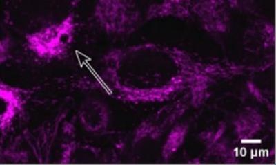
There are various biomedical markers present in cancerous tissues at higher concentrations than in healthy tissues—amino acids (building blocks of proteins), proteins, and enzymes. When excited with UV light, these markers light up and fluoresce in the UV and part of the visible spectrum, in a process called autofluorescence. “Imaging in the UV region has been limited and I would say that has been the biggest roadblock for making scientific progress,” explains Nie. “Now we have come up with this technology where we can image UV light with high sensitivity and can also distinguish small wavelength differences.”
This new imaging technology is enabling us to differentiate cancerous versus healthy cells and is opening up new and exciting applications beyond just health
Shuming Nie
Because cancer and healthy cells have different concentrations of markers and therefore different spectral signatures, the two classes of cells can be differentiated based on their fluorescence in the UV spectrum. The team evaluated their imaging device on its ability to discriminate cancer-related markers and found that is capable of differentiating between cancer and healthy cells with 99% confidence.
Gruev, Nie and their collaborative research team envision being able to use this sensor during surgery. One of the biggest challenges is knowing how much tissue to remove to ensure clear margins and such a sensor can help facilitate the decision-making process when a surgeon is removing a cancerous tumor.
“This new imaging technology is enabling us to differentiate cancerous versus healthy cells and is opening up new and exciting applications beyond just health,” Nie says. There are many other species besides butterflies capable of seeing in the UV, and having a way to detect that light will provide interesting opportunities for biologists to learn more about these species, such as their hunting and mating habits. Bringing the sensor underwater can help bring a greater understanding of that environment as well. While a lot of UV is absorbed by water, there is still enough that makes it through to have an impact and there are many animals underwater that also see and use UV light.
Source: The Grainger College of Engineering at University of Illinois Urbana-Champaign
06.11.2023