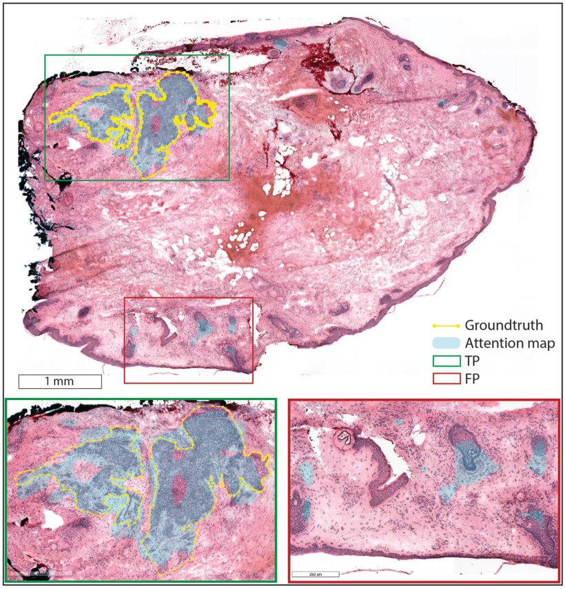
Image source: Geijs DJ, Hillen LM, Dooper S et al., Modern Pathology 2025 (CC BY 4.0)
News • Faster, more accurate treatment
Basal cell carcinoma: AI support for Mohs surgery
New tool supports faster, more accurate treatment of basal cell carcinoma during Mohs surgery
Dutch researchers have developed an AI tool that could transform how surgeons treat the most common form of cancer in the Netherlands: basal cell carcinoma. The system is designed to support Mohs surgery, a precise but time-consuming procedure, by speeding up tissue analysis and reducing reliance on specialized pathologists.
Basal cell carcinoma (BCC) is the most common form of cancer in the Netherlands. Though rarely life-threatening, if left untreated—particularly on the face—it can lead to disfigurement or loss of function. Mohs micrographic surgery (MMS) is the gold standard for treating BCC, as it allows surgeons to precisely remove cancerous tissue while sparing as much healthy skin as possible. However, the procedure is time-consuming and requires the expertise of trained dermatopathologists, who are not always available. To address this challenge, researchers from Radboudumc and Maastricht UMC+ have developed an AI-powered tool that supports the analysis of frozen tissue samples during Mohs surgery. Their study, published in Modern Pathology, demonstrates how artificial intelligence can assist surgeons in identifying cancerous regions more quickly and confidently—potentially reducing surgery times and improving patient care.
Recommended article

News • Basal cell carcinoma diagnostics
BCC: AI helps identify aggressive types
Dermatologists could benefit from a new algorithm that can help recognize patients with a highly aggressive form of basal cell carcinoma (BCC) of the face.
Instead of relying solely on a pathologist, the AI system functions as a virtual assistant, highlighting suspicious areas in tissue samples. By visually guiding the surgeon and marking possible tumour regions, the tool speeds up the analysis and reduces the risk of missing residual cancer cells.
While promising, the technology still requires further validation and regulatory approval before it can be used in clinical practice. Ongoing research will focus on ensuring its safety, reliability, and seamless integration into medical workflows. With these developments, AI-driven support tools could play an increasingly vital role in helping doctors manage rising cancer caseloads and deliver high-quality care more efficiently.
Source: Radboudumc
26.04.2025