News • High-resolution cell scans
AI spots cancer and viral infections at nanoscale precision
Scanning images of cells could lead to new diagnostic and monitoring strategies for disease
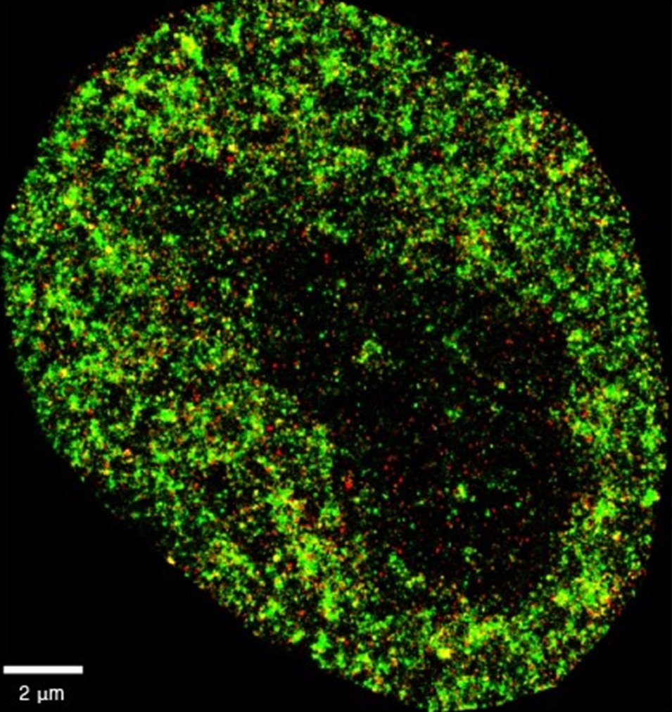
Image source: Center for Genomic Regulation; credit: Zhong Limei (CC BY-SA 4.0)
Researchers at the Centre for Genomic Regulation (CRG), the University of the Basque Country (UPV/EHU), Donostia International Physics Center (DIPC) and the Fundación Biofisica Bizkaia (FBB, located in Biofisika Institute) have developed an artificial intelligence which can differentiate cancer cells from normal cells, as well as detect the very early stages of viral infection inside cells. The findings, published in the journal Nature Machine Intelligence, pave the way for improved diagnostic techniques and new monitoring strategies for disease.
The tool, AINU (AI of the NUcleus), scans high-resolution images of cells. The images are obtained with a special microscopy technique called STORM, which creates a picture that captures many finer details than what regular microscopes can see. The high-definition snapshots reveal structures at nanoscale resolution.
A nanometre (nm) is one-billionth of a metre, and a strand of human hair is about 100,000nm wide. The AI can detect rearrangements inside cells as small as 20nm, or 5,000 times smaller than the width of a human hair. These alterations are too small and subtle for human observers to find with traditional methods alone. “The resolution of these images is powerful enough for our AI to recognise specific patterns and differences with remarkable accuracy, including changes in how DNA is arranged inside cells, helping spot alterations very soon after they occur. We think that, one day, this type of information can buy doctors valuable time to monitor disease, personalise treatments and improve patient outcomes,” says ICREA Research Professor Pia Cosma, co-corresponding author of the study and researcher at the Centre for Genomic Regulation in Barcelona.
AINU is a convolutional neural network, a type of AI specifically designed to analyse visual data like images. Examples of convolutional neural networks include AI tools which enables users to unlock smartphones with their face, or others used by self-driving cars to understand and navigate environments by recognising objects on the road. In medicine, convolutional neural networks are used to analyse medical images like mammograms or CT scans and identify signs of cancer that might be missed by the human eye. They can also help doctors detect abnormalities in MRI scans or X-ray images, helping make a faster and more accurate diagnosis.
Recommended article

Article • Technology overview
Artificial intelligence (AI) in healthcare
With the help of artificial intelligence, computers are to simulate human thought processes. Machine learning is intended to support almost all medical specialties. But what is going on inside an AI algorithm, what are its decisions based on? Can you even entrust a medical diagnosis to a machine? Clarifying these questions remains a central aspect of AI research and development.
AINU detects and analyses tiny structures inside cells at the molecular level. The researchers trained the model by feeding it with nanoscale-resolution images of the nucleus of many different types of cells in different states. The model learned to recognize specific patterns in cells by analysing how nuclear components are distributed and arranged in three-dimensional space. For example, cancer cells have distinct changes in their nuclear structure compared to normal cells, such as alterations to how their DNA is organised or the distribution of enzymes within the nucleus. After training, AINU could analyse new images of cell nuclei and classify them as cancerous or normal based on these features alone.
All our AI model needs to work is a sample that is stained with specific markers that highlight key nuclear features. As well as being easier and faster, it can accelerate stem cell research while contributing to the shift in reducing animal use in science
Davide Carnevali
The nanoscale resolution of the images enabled the AI detect changes in a cell's nucleus as soon as one hour after it was infected by the herpes simplex virus type-1. The model could detect the presence of the virus by finding slight differences in how tightly DNA is packed, which happens when a virus starts to alter the structure of the cell's nucleus. “Our method can detect cells that have been infected by a virus very soon after the infection starts. Normally, it takes time for doctors to spot an infection because they rely on visible symptoms or larger changes in the body. But with AINU, we can see tiny changes in the cell's nucleus right away,” says Ignacio Arganda-Carreras, co-corresponding author of the study and Ikerbasque Research Associate at UPV/EHU and affiliated with the FBB-Biofisika Institute and the DIPC in San Sebastián/Donostia.
“Researchers can use this technology to see how viruses affect cells almost immediately after they enter the body, which could help in developing better treatments and vaccines. hospitals and clinics, AINU could be used to quickly diagnose infections from a simple blood or tissue sample, making the process faster and more accurate,” adds Limei Zhong, co-first author of the study and researcher at the Guangdong Provincial People's Hospital (GDPH) in Guangzhou, China.
The researchers have to overcome important limitations before the technology is ready to be tested or deployed in a clinical setting. For example, STORM images can only be taken with specialized equipment normally only found in biomedical research labs. Setting up and maintaining the imaging systems required by the AI is a significant investment in both equipment and technical expertise.
Another constraint is that STORM imaging typically analyses only a few cells at a time. For diagnostic purposes, especially in clinical settings where speed and efficiency are crucial, doctors would need to capture many more numbers of cells in a single image to be able to detect or monitor a disease. “There are many rapid advances in the field of STORM imaging which mean that microscopes may soon be available in smaller or less specialized labs, and eventually, even in the clinic. The limitations of accessibility and throughput are more tractable problems than we previously thought and we hope to carry out preclinical experiments soon,” says Dr. Cosma.
Though clinical benefits might be years away, AINU is expected to accelerate scientific research in the short term. The researchers found the technology could identify stem cells with very high precision. Stem cells can develop into any type of cell in the body, an ability known as pluripotency. Pluripotent cells are studied for their potential in helping repair or replace damaged tissues.
AINU can make the process of detecting pluripotent cells quicker and more accurate, helping make stem cell therapies safer and more effective. “Current methods to detect high-quality stem cells rely on animal testing. However, all our AI model needs to work is a sample that is stained with specific markers that highlight key nuclear features. As well as being easier and faster, it can accelerate stem cell research while contributing to the shift in reducing animal use in science,” says Davide Carnevali, first author of the research and researcher at the CRG.
Source: Center for Genomic Regulation
27.08.2024