Image source: LiU/Emma Busk Winquist
News • Imaging white matter damage
Advanced MRI detects brain changes after Covid-19
Researchers at Linköping University have examined the brains of 16 patients previously hospitalised for Covid-19 with persisting symptoms. They have found differences in brain tissue structure between patients with persisting symptoms after Covid-19 and healthy people.
Their findings, published in the journal Brain Communications, can bring insights into the underlying mechanisms of persisting neurological problems after Covid-19.

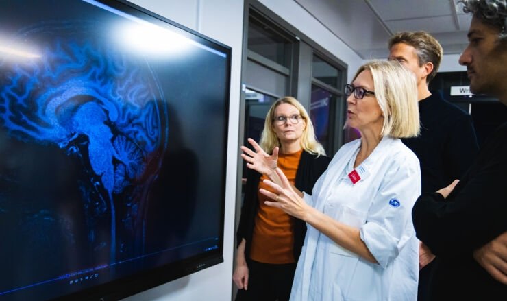
Image source: LiU
Several previous studies of persisting problems after Covid have involved MRI brain scanning. Although researchers have found differences compared with healthy brains, these differences are not specific to Covid-19.
“It can be frustrating for me as a doctor when I understand that the patients have problems, but I can’t find an explanation because there’s nothing in the MRI scan to explain it. To me, this underlines the importance of trying other examination technologies to understand what’s happening in the brain in patients with persisting symptoms after Covid-19,” says Ida Blystad, neuroradiologist in the Department of Radiology at Linköping University Hospital and researcher affiliated with the Department of Health, Medicine and Caring Sciences at Linköping University and the Centre for Medical Image Science and Visualization (CMIV).
In their current study, the researchers have therefore added a new type of MR imaging called advanced diffusion MRI. They were particularly interested in the brain’s white matter. This consists mainly of nerve axons and is very important for transporting signals between the different parts of the brain and the rest of the body.

Image source: LiU/Magnus Johansson
“Diffusion MRI is a very sensitive technology that allows changes in how the nerve axons are organised to be detected. This is one of the reasons why we wanted to use diffusion MRI to study the effects of Covid-19 on the brain that other imaging technologies might not pick up,” says Deneb Boito, doctoral student at the Department of Biomedical Engineering at Linköping University.
To get an idea of what diffusion MRI is, we can imagine a big city at night. Car headlights and rear lights shine like red and white strings of pearls on the most trafficked roads. We cannot see the road itself, but we understand that it is there, as the cars can easily move about right there. Similarly, doctors and researchers can get an insight into how the brain is constructed on a microscopic level through diffusion MRI. This technology builds on the fact that there is water everywhere in the brain moving in the tissue according to the law of least resistance. Water molecules move more easily along the neural pathways. By measuring the movement of water molecules through the neural pathways, researchers can indirectly infer the structure of neural pathways, just as we can indirectly understand that there is a motorway where there are many cars driving.
To me, these findings are a sign that we must investigate long-term effects of Covid-19 in the brain using more advanced MRI technology than conventional MRI
Ida Blystad
Healthcare usages of diffusion MRI include diagnosing stroke and planning brain surgery. In their current study, the researchers used a more advanced version of diffusion MRI. They examined 16 men who had been hospitalised for severe Covid-19 and who are participating in the Linköping Covid-19 Study (LinCos) at the Department of Rehabilitation Medicine in Linköping. They still had persisting symptoms after seven months. This group was compared with a group of healthy individuals without post-Covid symptoms who had not been hospitalised for Covid. The participants’ brains were examined with both conventional MRI and diffusion MRI.
“The two groups differ when it comes to brain white matter structure. This can be one of the causes of the neurological problems experienced by the group that had suffered from severe Covid-19. It’s a result that’s in line with other studies that have shown changes to the brain’s white matter. However, having examined only a small group of patients, we are cautious about drawing any major conclusions. With this technology, we’re not measuring the function of the brain, but its microstructure. To me, these findings are a sign that we must investigate long-term effects of Covid-19 in the brain using more advanced MRI technology than conventional MRI,” says Ida Blystad.
Recommended article
Article • Covid-19
Coronavirus update
Years after the first outbreak and spread of coronavirus SARS-CoV-2, its impact can still be felt in everyday life. Keep up-to-date with the latest research news, political developments, and background information on Covid-19.
There are several issues that the researchers want to study further. It appears, for instance, that white matter in different parts of the brain is affected in different ways, although it is too early to draw any conclusions as to what these differences mean. An upcoming study will investigate whether changes detected with diffusion MRI are in any way connected to brain activity, and how different parts of the brain communicate with each other through the brain white matter in patients suffering from post-Covid fatigue.
Another question is what happens over time. The MRI scan provides an image of the brain at that particular moment. As the participants were examined on one occasion only, it is not possible to know whether the differences between the two groups will disappear over time or whether they are permanent.
This research was funded by, among others, the Analytic Imaging Diagnostic Arena (AIDA), the ITEA/Vinnova project ASSIST, and the Wallenberg Center for Molecular Medicine at Linköping University.
Source: Linköping University
13.12.2023